5ZRS image
Entry Detail
PDB ID:
5ZRS
Keywords:
Title:
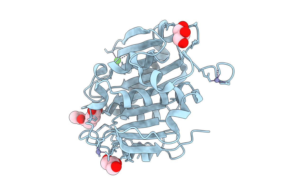
Crystal structure of PET-degrading cutinase Cut190 S176A/S226P/R228S mutant in monoethyl adipate bound state
Biological Source:
Source Organism:
Host Organism:
PDB Version:
Deposition Date:
2018-04-25
Release Date:
2018-09-12
Method Details:
Experimental Method:
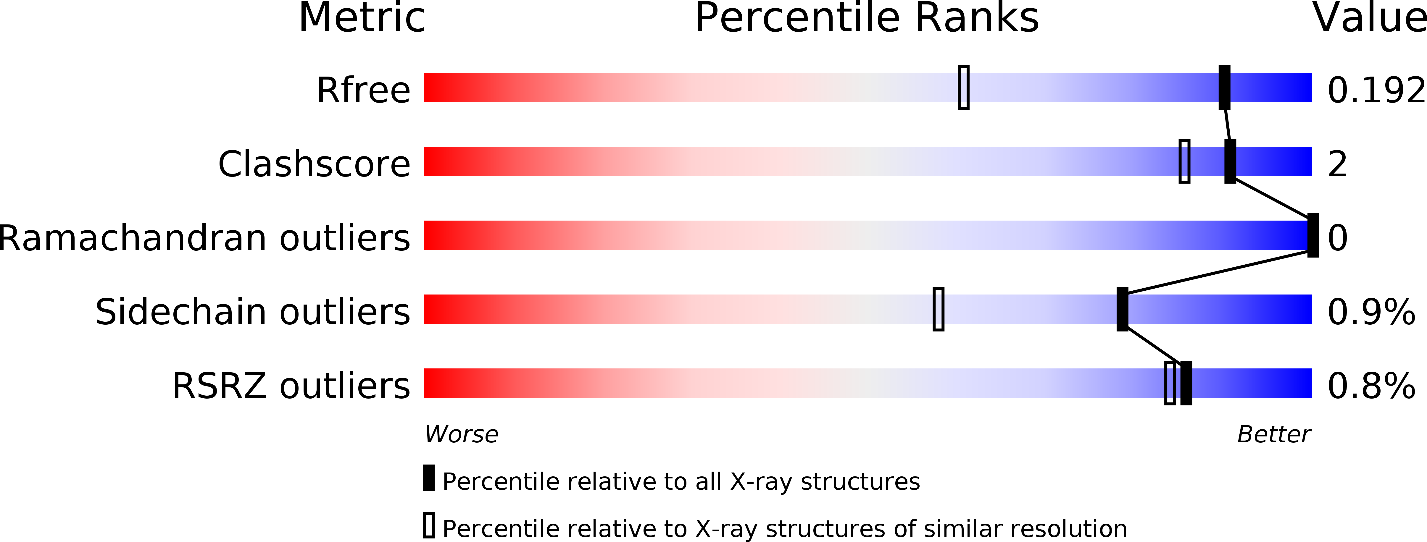
Resolution:
1.40 Å
R-Value Free:
0.19
R-Value Work:
0.16
R-Value Observed:
0.16
Space Group:
P 21 21 21
Macromolecular Entities
Polymer Type:polypeptide(L)
Description:Alpha/beta hydrolase family protein
Mutations:S176A, S226P, R228S
Chain IDs:A
Chain Length:265
Number of Molecules:1
Biological Source:Saccharomonospora viridis
Modified Residue
Compound ID Chain ID Parent Comp ID Details 2D Image
MHO A MET modified residue
Primary Citation
Structural Dynamics of the PET-Degrading Cutinase-like Enzyme from Saccharomonospora viridis AHK190 in Substrate-Bound States Elucidates the Ca2+-Driven Catalytic Cycle.
Biochemistry 57 5289 5300 (2018)
PMID: 30110540 DOI: 10.1021/acs.biochem.8b00624

Abstact

A cutinase-type polyesterase from Saccharomonospora viridis AHK190 (Cut190) has been shown to degrade the inner block of polyethylene terephthalate. A unique feature of Cut190 is that its function and stability are regulated by Ca2+ binding. Our previous crystal structure analysis of Cut190S226P showed that one Ca2+ binds to the enzyme, which induces large conformational changes in several loop regions to stabilize an open conformation [Miyakawa, T., et al. (2015) Appl. Microbiol. Biotechnol. 99, 4297]. In this study, to analyze the substrate recognition mechanism of Cut190, we determined the crystal structure of the inactive form of a Cut190 mutant, Cut190*S176A, in complex with calcium ions and/or substrates. We found that three calcium ions bind to Cut190*S176A, which is supported by analysis using native mass spectrometry experiments and 3D Reference Interaction Site Model calculations. The complex structures with the two substrates, monoethyl succinate and monoethyl adipate (engaged and open forms), presumably correspond to the pre- and post-reaction states, as the ester bond is close to the active site and pointing outward from the active site, respectively, for the two complexes. Ca2+ binding induces the pocket to open, enabling the substrate to access the pocket more easily. Molecular dynamics simulations suggest that a post-reaction state in the engaged form presumably exists between the experimentally observed forms, indicating that the substrate would be cleaved in the engaged form and then requires the enzyme to change to the open form to release the product, a process that Ca2+ can greatly accelerate.

Legend

Protein

Chemical

Disease

Primary Citation of related structures