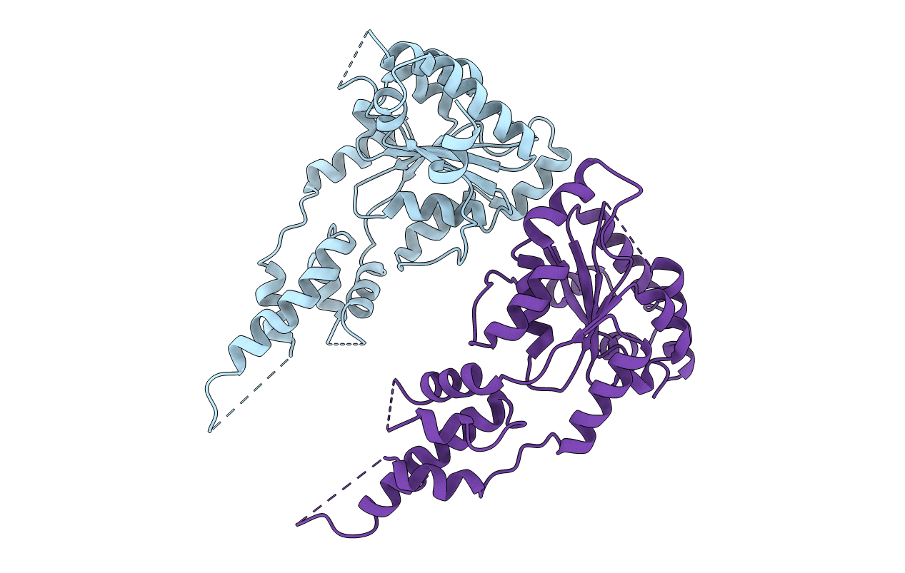
5ZQL image
Entry Detail
PDB ID:
5ZQL
Keywords:
Title:
crystal structure of human katanin AAA ATPase domain
Biological Source:
Source Organism:
Host Organism:
PDB Version:
Deposition Date:
2018-04-19
Release Date:
2019-02-27
Method Details:
Experimental Method:
Resolution:
3.01 Å
R-Value Free:
0.28
R-Value Work:
0.27
R-Value Observed:
0.27
Space Group:
P 43
Macromolecular Entities
Polymer Type:polypeptide(L)
Description:Katanin p60 ATPase-containing subunit A1
Mutations:E309Q
Chain IDs:A (auth: B), B (auth: A)
Chain Length:309
Number of Molecules:2
Biological Source:Homo sapiens
Ligand Molecules
Primary Citation
Structural and Molecular Basis for Katanin-Mediated Severing of Glutamylated Microtubules.
Cell Rep 26 1357 1367.e5 (2019)
PMID: 30699360 DOI: 10.1016/j.celrep.2019.01.020

Abstact

Katanin was the first microtubule (MT)-severing enzyme discovered, but how katanin executes MT severing remains poorly understood. Here, we report X-ray crystal structures of the apo and ATPγS-bound states of the catalytic AAA domain of human katanin p60 at 3.0 and 2.9 Å resolution, respectively. Comparison of the two structures reveals conformational changes induced by ATP binding and how such changes ensure hexamer stability. Moreover, we uncover structural details of pore loops (PLs) and show that Arg283, a residue unique to katanin among MT-severing enzymes, protrudes from PL1 and lines the entry of the catalytic pore. Functional studies suggest that PL1 and Arg283 play essential roles in the recognition and remodeling of the glutamylated, C-terminal tubulin tail and regulation of axon growth. In addition, domain-swapping experiments in katanin and spastin suggest that the non-homologous N-terminal region, which contains the MT-interacting and trafficking domain and a linker, confers specificity to the severing process.

Legend

Protein

Chemical

Disease

Primary Citation of related structures