5ZN8 image
Deposition Date 2018-04-08
Release Date 2018-04-25
Last Version Date 2023-11-22
Entry Detail
PDB ID:
5ZN8
Title:
Crystal structure of nicotinamidase PncA from Bacillus subtilis
Biological Source:
Source Organism:
Host Organism:
Method Details:
Experimental Method:
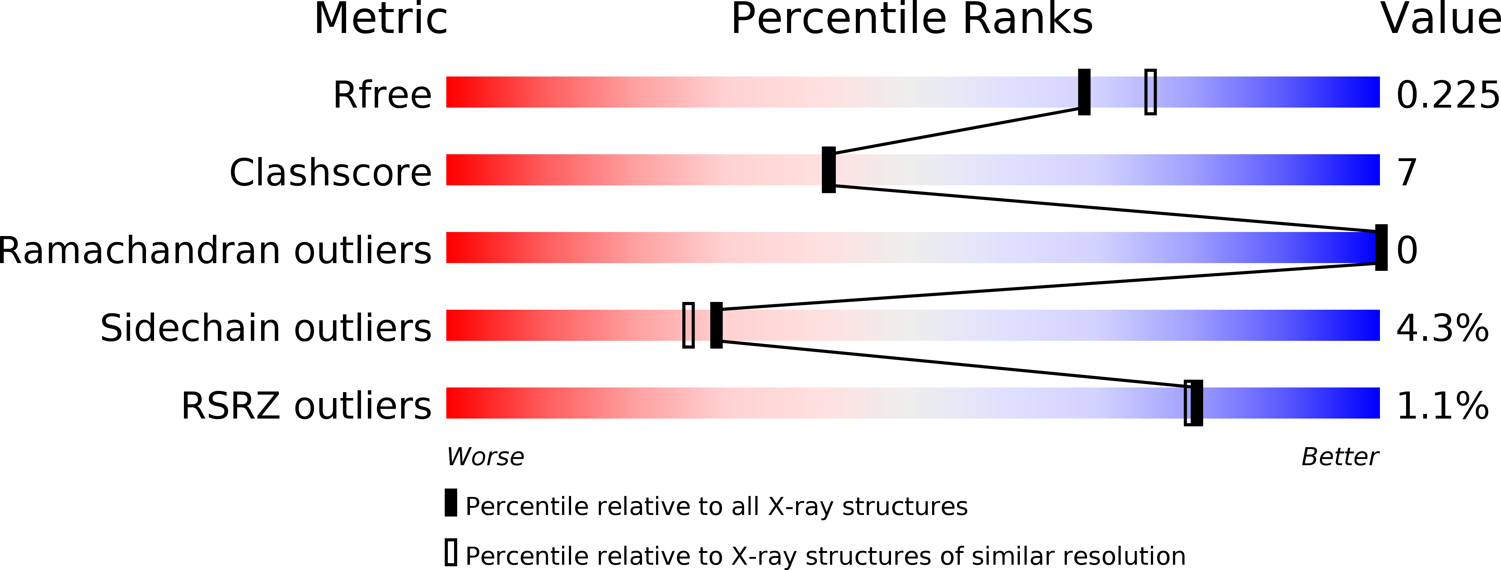
Resolution:
2.00 Å
R-Value Free:
0.22
R-Value Work:
0.15
R-Value Observed:
0.15
Space Group:
P 62
Macromolecular Entities
Polymer Type:polypeptide(L)
Molecule:Isochorismatase
Mutagens:F5L
Chain IDs:A, B
Chain Length:183
Number of Molecules:2
Biological Source:Bacillus subtilis
Ligand Molecules
Primary Citation
Crystal structure of the nicotinamidase/pyrazinamidase PncA from Bacillus subtilis.
Biochem.Biophys.Res.Commun. 503 2906 2911 (2018)
PMID: 30107912 DOI: 10.1016/j.bbrc.2018.08.067

Abstact

The nicotinamidase/pyrazinamidase PncA is a member of a large family of hydrolase enzymes that catalyze the deamination of nicotinamide to nicotinic acid. PncA also functions as a pyrazinamidase in a wide variety of eubacteria and is an essential coenzyme in many cellular redox reactions in living systems. We report the crystal structure of substrate-free PncA from Bacillus subtilis (BsPncA) at 2.0 Å resolution to improve our understanding of the PncA family. The structure of BsPncA consists of an α/β domain and a subdomain. The subdomain of BsPncA has a different conformation than that of PncA enzymes from other organisms. The B-factor analysis revealed a rigid structure of the α/β domain, while the subdomain is highly flexible. Both dimers and tetramers were observed in BsPncA protein crystals, but only dimers were observed in solution. Our results provide useful information that will further enhance our understanding of the molecular functions of PncA family members.

Legend

Protein

Chemical

Disease

Primary Citation of related structures
Feedback Form
Name
Email
Institute
Feedback