Deposition Date
2018-04-05
Release Date
2018-10-17
Last Version Date
2023-11-22
Entry Detail
PDB ID:
5ZMS
Keywords:
Title:
Crystal structure of Zika NS3 protease in complex with 4-guanidinomethyl-phenylacetyl-Lys-Lys-Arg-H
Biological Source:
Source Organism(s):
Zika virus (Taxon ID: 64320)
synthetic construct (Taxon ID: 32630)
synthetic construct (Taxon ID: 32630)
Expression System(s):
Method Details:
Experimental Method:
Resolution:
1.80 Å
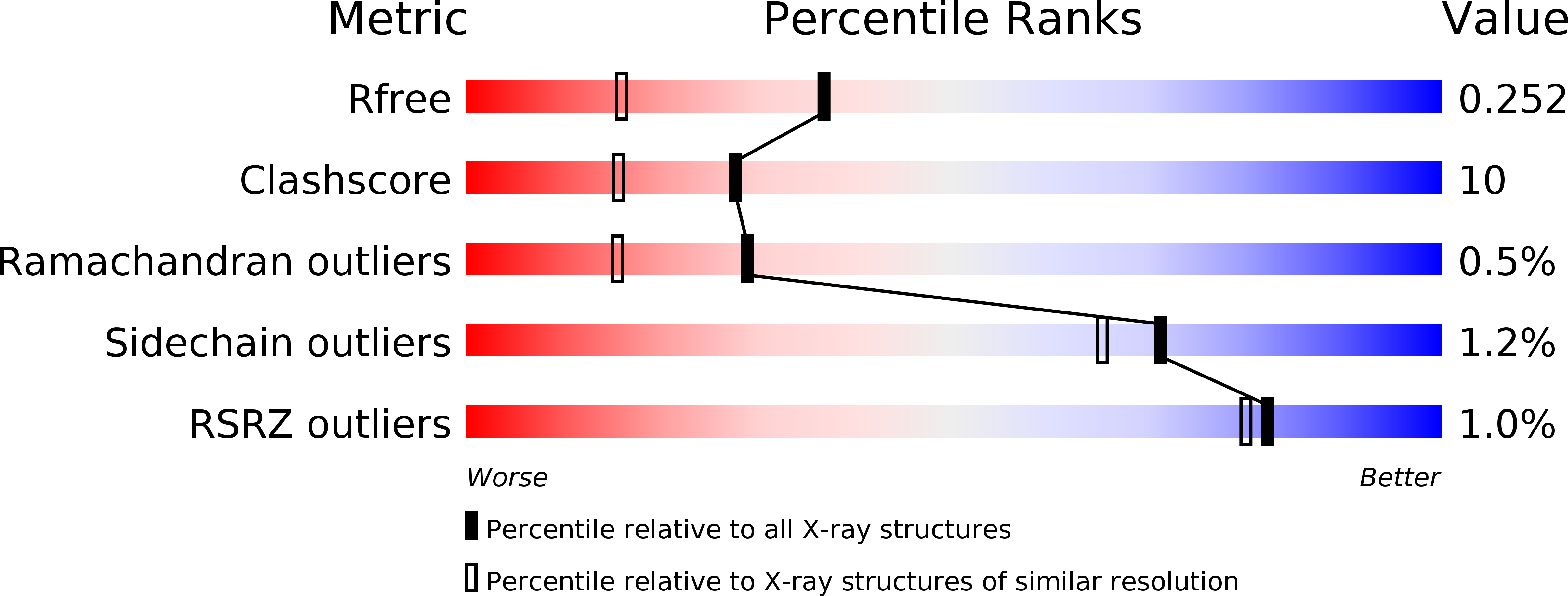
R-Value Free:
0.25
R-Value Work:
0.20
R-Value Observed:
0.21
Space Group:
P 21 21 21