
Deposition Date
2018-03-14
Release Date
2018-08-01
Last Version Date
2024-10-09
Entry Detail
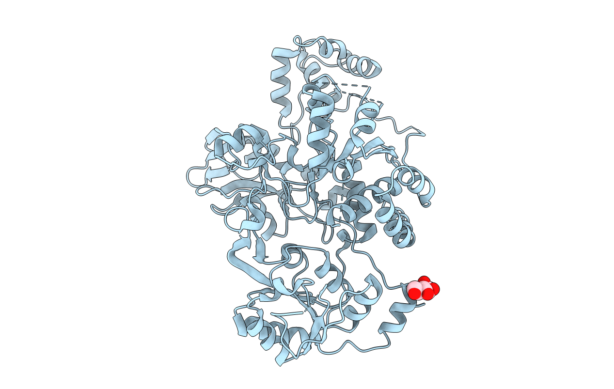
PDB ID:
5ZIB
Keywords:
Title:
Crystal structure of human GnT-V luminal domain in apo form
Biological Source:
Source Organism(s):
Homo sapiens (Taxon ID: 9606)
Expression System(s):
Method Details:
Experimental Method:
Resolution:
1.90 Å
R-Value Free:
0.22
R-Value Work:
0.18
R-Value Observed:
0.19
Space Group:
P 65 2 2
