5ZCK image
Deposition Date 2018-02-18
Release Date 2018-04-18
Last Version Date 2023-11-22
Entry Detail
PDB ID:
5ZCK
Keywords:
Title:
Structure of the RIP3 core region
Biological Source:
Source Organism:
Homo sapiens (Taxon ID: 9606)
Method Details:
Experimental Method:
Resolution:
1.27 Å
R-Value Free:
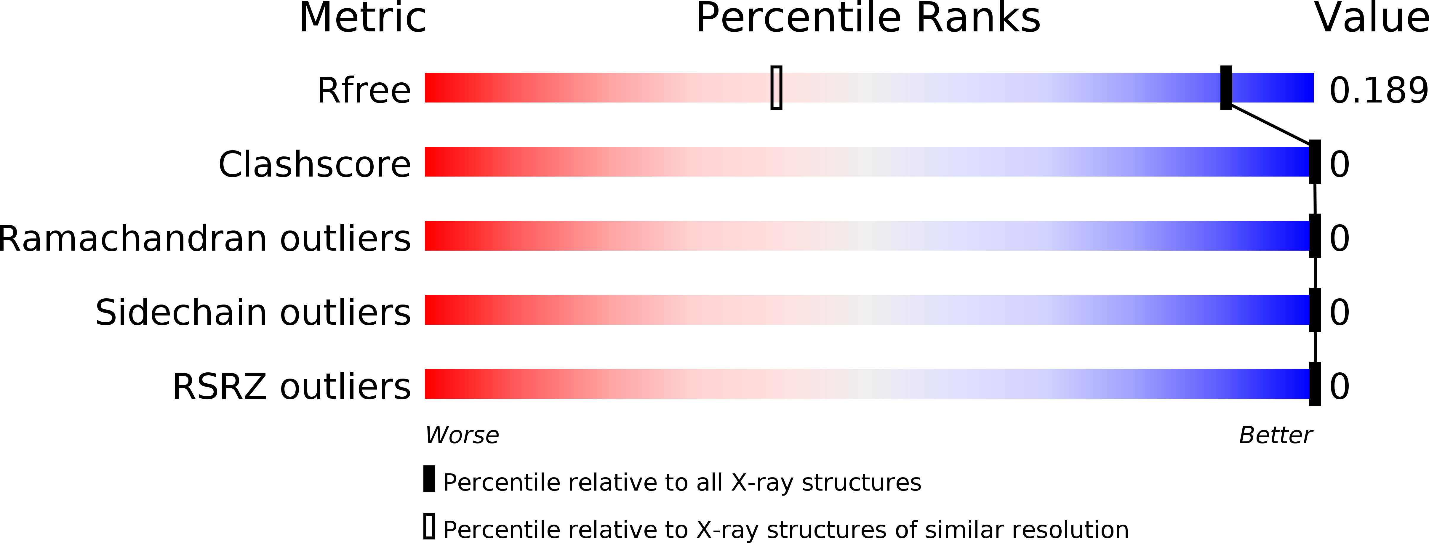
0.15
R-Value Work:
0.13
R-Value Observed:
0.13
Space Group:
P 21 21 21
Macromolecular Entities
Polymer Type:polypeptide(L)
Molecule:peptide from Receptor-interacting serine/threonine-protein kinase 3
Gene (Uniprot):RIPK3
Chain IDs:A
Chain Length:4
Number of Molecules:1
Biological Source:Homo sapiens
Ligand Molecules
Primary Citation
The Structure of the Necrosome RIPK1-RIPK3 Core, a Human Hetero-Amyloid Signaling Complex.
Cell 173 1244 1253.e10 (2018)
PMID: 29681455 DOI: 10.1016/j.cell.2018.03.032

Abstact

The RIPK1-RIPK3 necrosome is an amyloid signaling complex that initiates TNF-induced necroptosis, serving in human immune defense, cancer, and neurodegenerative diseases. RIPK1 and RIPK3 associate through their RIP homotypic interaction motifs with consensus sequences IQIG (RIPK1) and VQVG (RIPK3). Using solid-state nuclear magnetic resonance, we determined the high-resolution structure of the RIPK1-RIPK3 core. RIPK1 and RIPK3 alternately stack (RIPK1, RIPK3, RIPK1, RIPK3, etc.) to form heterotypic β sheets. Two such β sheets bind together along a compact hydrophobic interface featuring an unusual ladder of alternating Ser (from RIPK1) and Cys (from RIPK3). The crystal structure of a four-residue RIPK3 consensus sequence is consistent with the architecture determined by NMR. The RIPK1-RIPK3 core is the first detailed structure of a hetero-amyloid and provides a potential explanation for the specificity of hetero- over homo-amyloid formation and a structural basis for understanding the mechanisms of signal transduction.

Legend

Protein

Chemical

Disease

Primary Citation of related structures