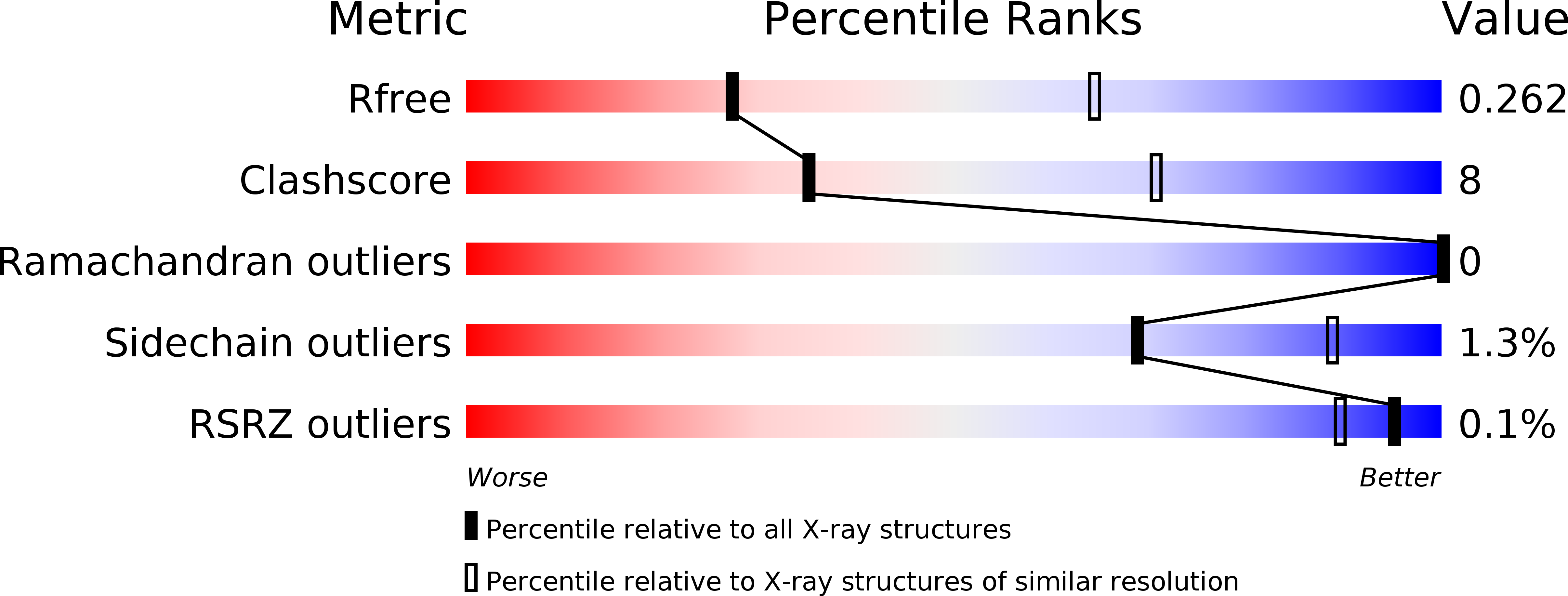
5ZC3 image
Deposition Date 2018-02-14
Release Date 2018-08-15
Last Version Date 2024-10-30
Entry Detail
PDB ID:
5ZC3
Title:
The Crystal Structure of PcRxLR12
Biological Source:
Source Organism:
Method Details:
Experimental Method:
Resolution:
3.01 Å
R-Value Free:
0.26
R-Value Work:
0.20
R-Value Observed:
0.21
Space Group:
P 21 21 21
Macromolecular Entities
Polymer Type:polypeptide(L)
Molecule:RxLR effector
Chain IDs:A (auth: B), B (auth: A)
Chain Length:472
Number of Molecules:2
Biological Source:Phytophthora capsici
Ligand Molecules
Primary Citation
Crystal structure of the RxLR effector PcRxLR12 from Phytophthora capsici
Biochem. Biophys. Res. Commun. 503 1830 1835 (2018)
PMID: 30077372 DOI: 10.1016/j.bbrc.2018.07.121

Abstact

RxLR genes are a prominent class of effectors in oomycetes, and almost half of these proteins contain a conserved sequence motif termed the WY domain, that may exist singly, or as divergent tandem repeats in different effectors. Here we describe the crystal structure of PcRxLR12 (63-488) from Phytophthora capsici at 3.0 Å resolution. The structure consists of five tandemly arrayed WY-domains linked to each other by short connecting helices. Superposition of the WY-2 domain on the other four domains of PcRxLR12, show that the first α-helix termed the K motif, and Loop 3 which connects α3 and α4 are the key regions of structural divergence between the WY domains. A similar pattern was observed when WY-2 was superposed on the 11 WY domains from other oomycete effectors. We also note that an added connecting helix between WY domains in some RXLR effectors, ensures that the WY domains are oriented in the same direction.

Legend

Protein

Chemical

Disease

Primary Citation of related structures