5ZC0 image
Entry Detail
PDB ID:
5ZC0
Title:
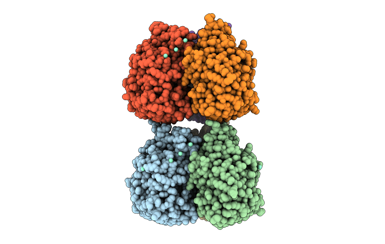
Crystal structure of Xenopus embryonic epidermal lectin in complex with Samarium ions
Biological Source:
Source Organism:
Host Organism:
PDB Version:
Deposition Date:
2018-02-14
Release Date:
2018-06-06
Method Details:
Experimental Method:
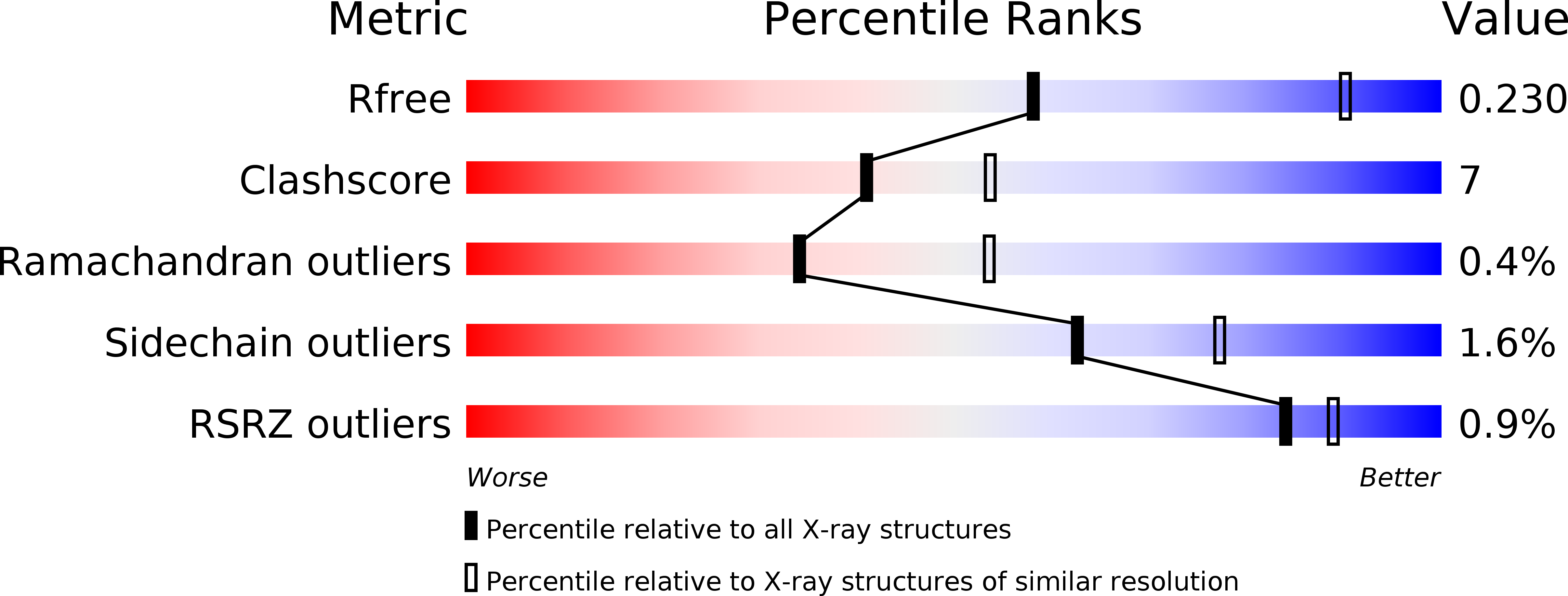
Resolution:
2.75 Å
R-Value Free:
0.23
R-Value Work:
0.19
R-Value Observed:
0.19
Space Group:
P 1 21 1
Macromolecular Entities
Polymer Type:polypeptide(L)
Description:Xenopus Embryonic Epidermal Lectin
Chain IDs:A, B, C, D, E, F
Chain Length:289
Number of Molecules:6
Biological Source:Xenopus laevis
Primary Citation
Structural stabilities of calcium proteins: Human intelectin-1 and frog lectin XEEL
J. Inorg. Biochem. 185 86 102 (2018)
PMID: 29807191 DOI: 10.1016/j.jinorgbio.2018.04.021

Abstact

We extend our study of the structural stability of helical and nonhelical regions in chain A of human intelectin-1 to include a second human intelectin (4WMY) and the frog protein "Xenopus embryonic epidermal lectin" (XEEL). These unique lectins have been shown to recognize carbohydrate residues found exclusively in microbes, thus they could potentially be developed into novel microbe detection and sequestration tools. We believe that by studying the structural stability of these proteins we can provide insights on their biological role and activities. Using a geometrical model introduced previously, we perform computational analyses of protein crystal structures that quantify the resiliency of the native state to steric perturbations. Based on these analyses, we conclude that differences in the resiliency of the human and frog proteins can be attributed primarily to differences in non-helical regions and to residues near Ca ions. Since these differences are particularly pronounced in the vicinity of the ligand binding site, they provide an explanation for the finding that human intelectin-1 has a higher affinity for a ligand than XEEL. We also present data on conserved and position-equivalent pairs of residues in 4WMY and XEEL. We identify residue pairs as well as regions in which the influence of neighboring residues is nearly uniform as the parent protein denatures. Since the structural signatures are conserved, this identification provides a basis for understanding why both proteins exhibit trimeric structures despite poor sequence conservation at the interface.

Legend

Protein

Chemical

Disease

Primary Citation of related structures