5Z86 image
Entry Detail
PDB ID:
5Z86
Keywords:
Title:
azide-bound cytochrome c oxidase structure determined using the crystals exposed to 20 mM azide solution for 3 days
Biological Source:
Source Organism:
PDB Version:
Deposition Date:
2018-01-31
Release Date:
2018-08-15
Method Details:
Experimental Method:
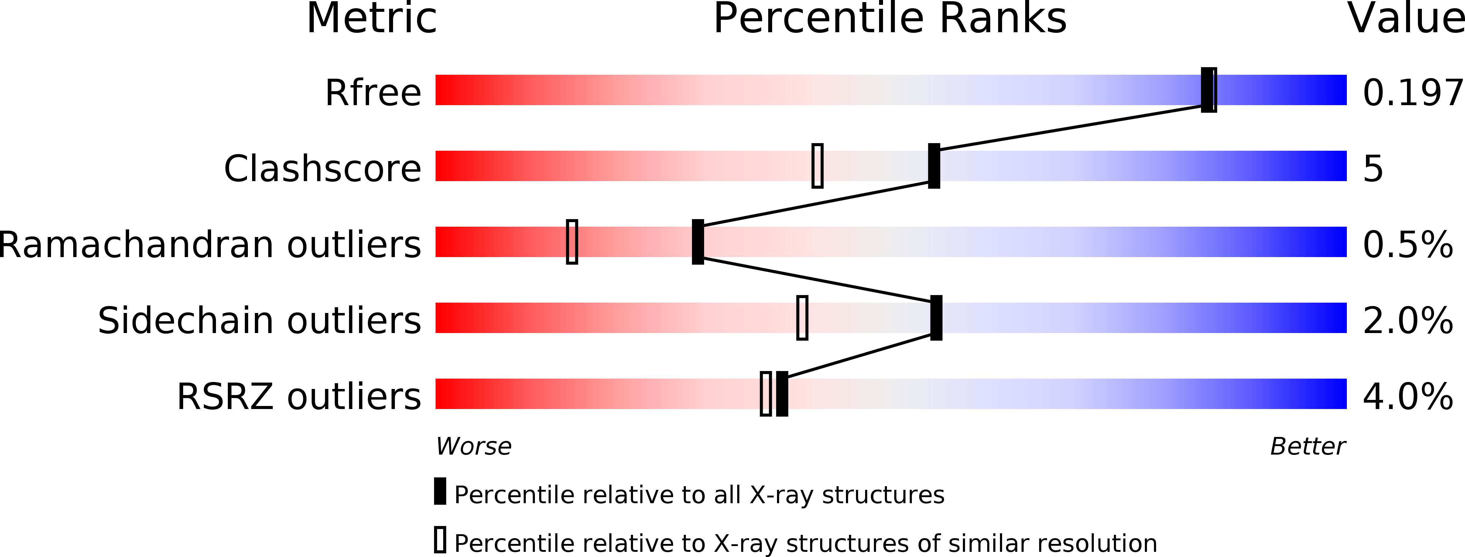
Resolution:
1.85 Å
R-Value Free:
0.19
R-Value Work:
0.18
R-Value Observed:
0.18
Space Group:
P 21 21 21
Macromolecular Entities
Polymer Type:polypeptide(L)
Description:Cytochrome c oxidase subunit 1
Chain IDs:A, N
Chain Length:514
Number of Molecules:2
Biological Source:Bos taurus
Polymer Type:polypeptide(L)
Description:Cytochrome c oxidase subunit 2
Chain IDs:B, O
Chain Length:227
Number of Molecules:2
Biological Source:Bos taurus
Polymer Type:polypeptide(L)
Description:Cytochrome c oxidase subunit 3
Chain IDs:C, P
Chain Length:261
Number of Molecules:2
Biological Source:Bos taurus
Polymer Type:polypeptide(L)
Description:Cytochrome c oxidase subunit 4 isoform 1, mitochondrial
Chain IDs:D, Q
Chain Length:147
Number of Molecules:2
Biological Source:Bos taurus
Polymer Type:polypeptide(L)
Description:Cytochrome c oxidase subunit 5A, mitochondrial
Chain IDs:E, R
Chain Length:109
Number of Molecules:2
Biological Source:Bos taurus
Polymer Type:polypeptide(L)
Description:Cytochrome c oxidase subunit 5B, mitochondrial
Chain IDs:F, S
Chain Length:98
Number of Molecules:2
Biological Source:Bos taurus
Polymer Type:polypeptide(L)
Description:Cytochrome c oxidase subunit 6A2, mitochondrial
Chain IDs:G, T
Chain Length:85
Number of Molecules:2
Biological Source:Bos taurus
Polymer Type:polypeptide(L)
Description:Cytochrome c oxidase subunit 6B1
Chain IDs:H, U
Chain Length:85
Number of Molecules:2
Biological Source:Bos taurus
Polymer Type:polypeptide(L)
Description:Cytochrome c oxidase subunit 6C
Chain IDs:I, V
Chain Length:73
Number of Molecules:2
Biological Source:Bos taurus
Polymer Type:polypeptide(L)
Description:Cytochrome c oxidase subunit 7A1, mitochondrial
Chain IDs:J, W
Chain Length:59
Number of Molecules:2
Biological Source:Bos taurus
Polymer Type:polypeptide(L)
Description:Cytochrome c oxidase subunit 7B, mitochondrial
Chain IDs:K, X
Chain Length:56
Number of Molecules:2
Biological Source:Bos taurus
Polymer Type:polypeptide(L)
Description:Cytochrome c oxidase subunit 7C, mitochondrial
Chain IDs:L, Y
Chain Length:47
Number of Molecules:2
Biological Source:Bos taurus
Polymer Type:polypeptide(L)
Description:Cytochrome c oxidase subunit 8B, mitochondrial
Chain IDs:M, Z
Chain Length:46
Number of Molecules:2
Biological Source:Bos taurus
Modified Residue
Compound ID Chain ID Parent Comp ID Details 2D Image
FME A MET modified residue
SAC I SER modified residue
TPO G THR modified residue
Primary Citation
X-ray structural analyses of azide-bound cytochromecoxidases reveal that the H-pathway is critically important for the proton-pumping activity.
J. Biol. Chem. 293 14868 14879 (2018)
PMID: 30077971 DOI: 10.1074/jbc.RA118.003123

Abstact

Cytochrome c oxidase (CcO) is the terminal oxidase of cellular respiration, reducing O2 to water and pumping protons. X-ray structural features have suggested that CcO pumps protons via a mechanism involving electrostatic repulsions between pumping protons in the hydrogen-bond network of a proton-conducting pathway (the H-pathway) and net positive charges created upon oxidation of an iron site, heme a (Fe a2+), for reduction of O2 at another iron site, heme a3 (Fe a32+). The protons for pumping are transferred to the hydrogen-bond network from the N-side via the water channel of the H-pathway. Back-leakage of protons to the N-side is thought to be blocked by closure of the water channel. To experimentally test this, we examined X-ray structures of the azide-bound, oxidized bovine CcO and found that an azide derivative (N3--Fe a33+, CuB2+-N3-) induces a translational movement of the heme a3 plane. This was accompanied by opening of the water channel, revealing that Fe a3 and the H-pathway are tightly coupled. The channel opening in the oxidized state is likely to induce back-leakage of pumping protons, which lowers the proton level in the hydrogen-bond network during enzymatic turnover. The proton level decrease weakens the electron affinity of Fe a , if Fe a electrostatically interacts with protons in the hydrogen-bond network. The previously reported azide-induced redox-potential decrease in Fe a supports existence of the electrostatic interaction. In summary, our results indicate that the H-pathway is critical for CcO's proton-pumping function.

Legend

Protein

Chemical

Disease

Primary Citation of related structures