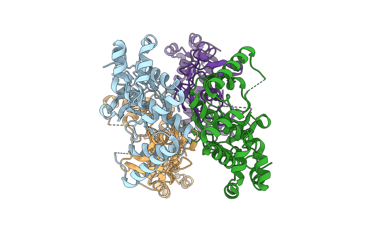
5Z5M image
Entry Detail
PDB ID:
5Z5M
Keywords:
Title:
Crystal structure of (S)-allantoin synthase
Biological Source:
PDB Version:
Deposition Date:
2018-01-18
Release Date:
2018-11-21
Method Details:
Experimental Method:
Resolution:
1.85 Å
R-Value Free:
0.23
R-Value Work:
0.19
R-Value Observed:
0.20
Space Group:
P 1 21 1
Macromolecular Entities
Polymer Type:polypeptide(L)
Description:Predicted protein
Chain IDs:A, B, C, D
Chain Length:299
Number of Molecules:4
Biological Source:Phaeodactylum tricornutum CCAP 1055/1
Primary Citation
Diatom Allantoin Synthase Provides Structural Insights into Natural Fusion Protein Therapeutics.
ACS Chem. Biol. 13 2237 2246 (2018)
PMID: 29874034 DOI: 10.1021/acschembio.8b00404

Abstact

Humans have lost the ability to convert urate into the more soluble allantoin with the evolutionary inactivation of three enzymes of the uricolytic pathway. Restoration of this function through enzyme replacement therapy can treat severe hyperuricemia and Lesch-Nyhan disease. Through a genomic exploration of natural gene fusions, we found that plants and diatoms independently evolved a fusion protein (allantoin synthase) complementing two human pseudogenes. The 1.85-Å-resolution crystal structure of allantoin synthase from the diatom Phaeodactylum tricornutum provides a rationale for the domain combinations observed in the metabolic pathway, suggesting that quaternary structure is key to the evolutionary success of protein domain fusions. Polyethylene glycol (PEG) conjugation experiments indicate that a PEG-modified form of the natural fusion protein provides advantages over separate enzymes in terms of activity maintenance and manufacturing of the bioconjugate. These results suggest that the combination of different activities in a single molecular unit can simplify the production and chemical modification of recombinant proteins for multifunctional enzyme therapy.

Legend

Protein

Chemical

Disease

Primary Citation of related structures