Deposition Date
2018-01-16
Release Date
2018-11-28
Last Version Date
2024-10-30
Entry Detail
PDB ID:
5Z51
Keywords:
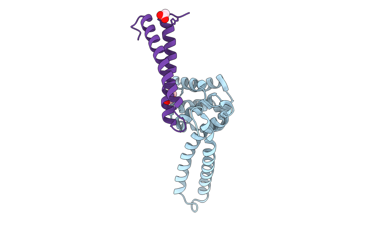
Title:
Helicase binding domain of primase from Mycobacterium tuberculosis
Biological Source:
Source Organism(s):
Mycobacterium tuberculosis H37Rv (Taxon ID: 83332)
Expression System(s):
Method Details:
Experimental Method:
Resolution:
1.58 Å
R-Value Free:
0.20
R-Value Work:
0.18
R-Value Observed:
0.18
Space Group:
P 21 21 2