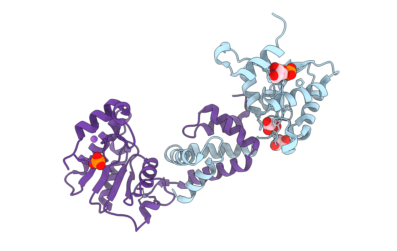
5Z2V image
Entry Detail
PDB ID:
5Z2V
Title:
Crystal structure of RecR from Pseudomonas aeruginosa PAO1
Biological Source:
PDB Version:
Deposition Date:
2018-01-04
Release Date:
2019-01-02
Method Details:
Experimental Method:
Resolution:
2.20 Å
R-Value Free:
0.21
R-Value Work:
0.17
R-Value Observed:
0.18
Space Group:
P 61 2 2
Macromolecular Entities
Polymer Type:polypeptide(L)
Description:Recombination protein RecR
Chain IDs:A, B
Chain Length:198
Number of Molecules:2
Biological Source:Pseudomonas aeruginosa (strain ATCC 15692 / DSM 22644 / CIP 104116 / JCM 14847 / LMG 12228 / 1C / PRS 101 / PAO1)
Primary Citation
Crystal structure of RecR, a member of the RecFOR DNA-repair pathway, from Pseudomonas aeruginosa PAO1.
Acta Crystallogr F Struct Biol Commun 74 222 230 (2018)
PMID: 29633970 DOI: 10.1107/S2053230X18003503

Abstact

DNA damage is usually lethal to all organisms. Homologous recombination plays an important role in the DNA damage-repair process in prokaryotic organisms. Two pathways are responsible for homologous recombination in Pseudomonas aeruginosa: the RecBCD pathway and the RecFOR pathway. RecR is an important regulator in the RecFOR homologous recombination pathway in P. aeruginosa. It forms complexes with RecF and RecO that can facilitate the loading of RecA onto ssDNA in the RecFOR pathway. Here, the crystal structure of RecR from P. aeruginosa PAO1 (PaRecR) is reported. PaRecR crystallizes in space group P6122, with two monomers per asymmetric unit. Analytical ultracentrifugation data show that PaRecR forms a stable dimer, but can exist as a tetramer in solution. The crystal structure shows that dimeric PaRecR forms a ring-like tetramer architecture via crystal symmetry. The presence of a ligand in the Walker B motif of one RecR subunit suggests a putative nucleotide-binding site.

Legend

Protein

Chemical

Disease

Primary Citation of related structures