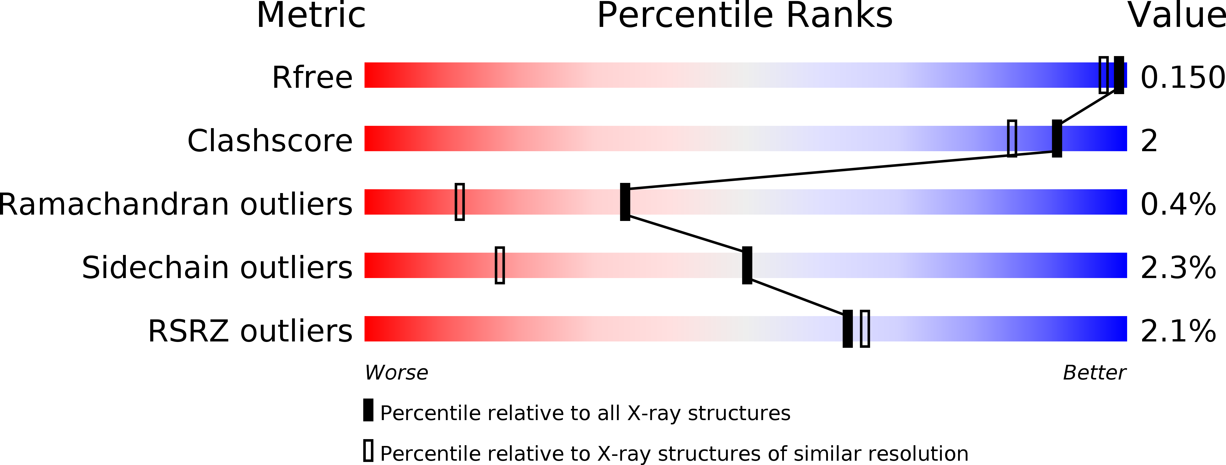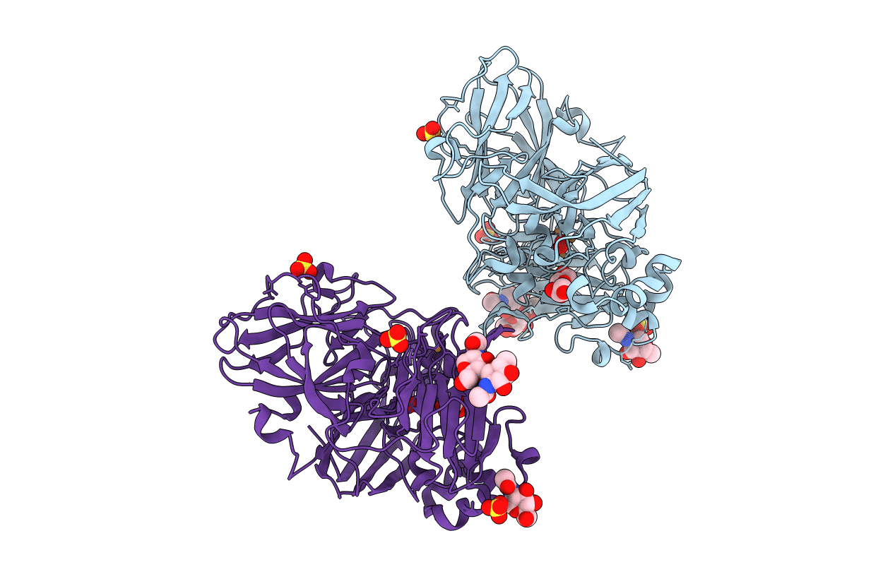
5Z1X image
Deposition Date 2017-12-28
Release Date 2018-09-05
Last Version Date 2024-11-06
Entry Detail
PDB ID:
5Z1X
Keywords:
Title:
Crystal Structure of Laccase from Cerrena sp. RSD1
Biological Source:
Source Organism:
Method Details:
Experimental Method:
Resolution:
1.38 Å
R-Value Free:
0.14
R-Value Work:
0.12
R-Value Observed:
0.12
Space Group:
P 21 21 21
Macromolecular Entities
Polymer Type:polypeptide(L)
Molecule:Laccase
Chain IDs:A, B
Chain Length:495
Number of Molecules:2
Biological Source:Cerrena sp. RSD1
Modified Residue
Compound ID Chain ID Parent Comp ID Details 2D Image
NAG C NAG -D
Primary Citation
Kinetic analysis and structural studies of a high-efficiency laccase fromCerrenasp. RSD1.
FEBS Open Bio 8 1230 1246 (2018)
PMID: 30087829 DOI: 10.1002/2211-5463.12459

Abstact

UNLABELLED A high-efficiency laccase, DLac, was isolated from Cerrena sp. RSD1. The kinetic studies indicate that DLac is a diffusion-limited enzyme. The crystal structure of DLac was determined to atomic resolution, and its overall structure shares high homology to monomeric laccases, but displays unique substrate-binding loops from those in other laccases. The substrate-binding residues with small side chain and the short substrate-binding loop IV broaden the substrate-binding cavity and may facilitate large substrate diffusion. Unlike highly glycosylated fungal laccases, the less-glycosylated DLac contains one highly conserved glycosylation site at N432 and an unique glycosylation site at N468. The N-glycans stabilize the substrate-binding loops and the protein structure, and the first N-acetylglucosamine is crucial for the catalytic efficiency. Additionally, a fivefold increase in protein yield is achieved via the submerged culture method for industrial applications. DATABASE The atomic coordinates of the structure of DLac from Cerrena sp. RSD1 and structural factors have been deposited in the RCSB Protein Data Bank (PDB ID: 5Z1X).

Legend

Protein

Chemical

Disease

Primary Citation of related structures