Deposition Date
2017-12-27
Release Date
2018-10-17
Last Version Date
2023-11-22
Entry Detail
PDB ID:
5Z1N
Keywords:
Title:
Crystal structure of C terminal region of G-protein interacting protein 1 (Gip1) from Dictyostelium discoideum
Biological Source:
Source Organism(s):
Dictyostelium discoideum (Taxon ID: 44689)
Expression System(s):
Method Details:
Experimental Method:
Resolution:
1.95 Å
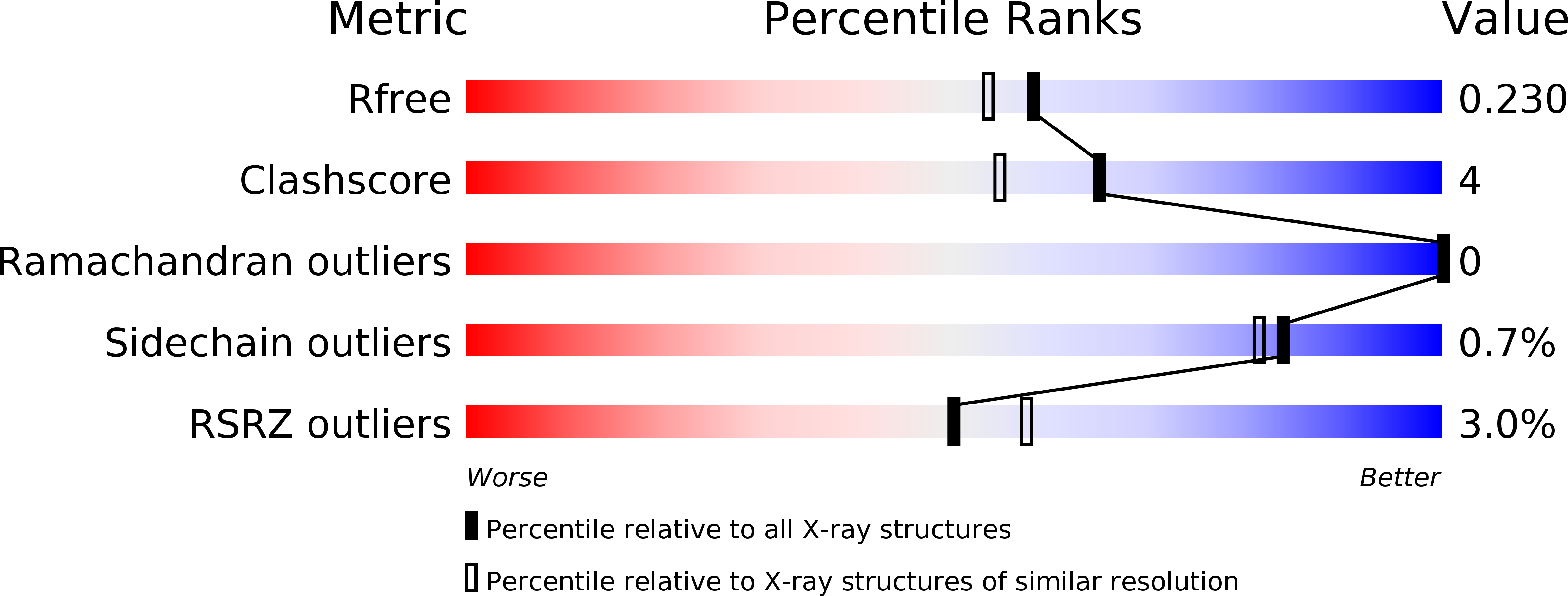
R-Value Free:
0.23
R-Value Work:
0.18
R-Value Observed:
0.18
Space Group:
P 21 21 21