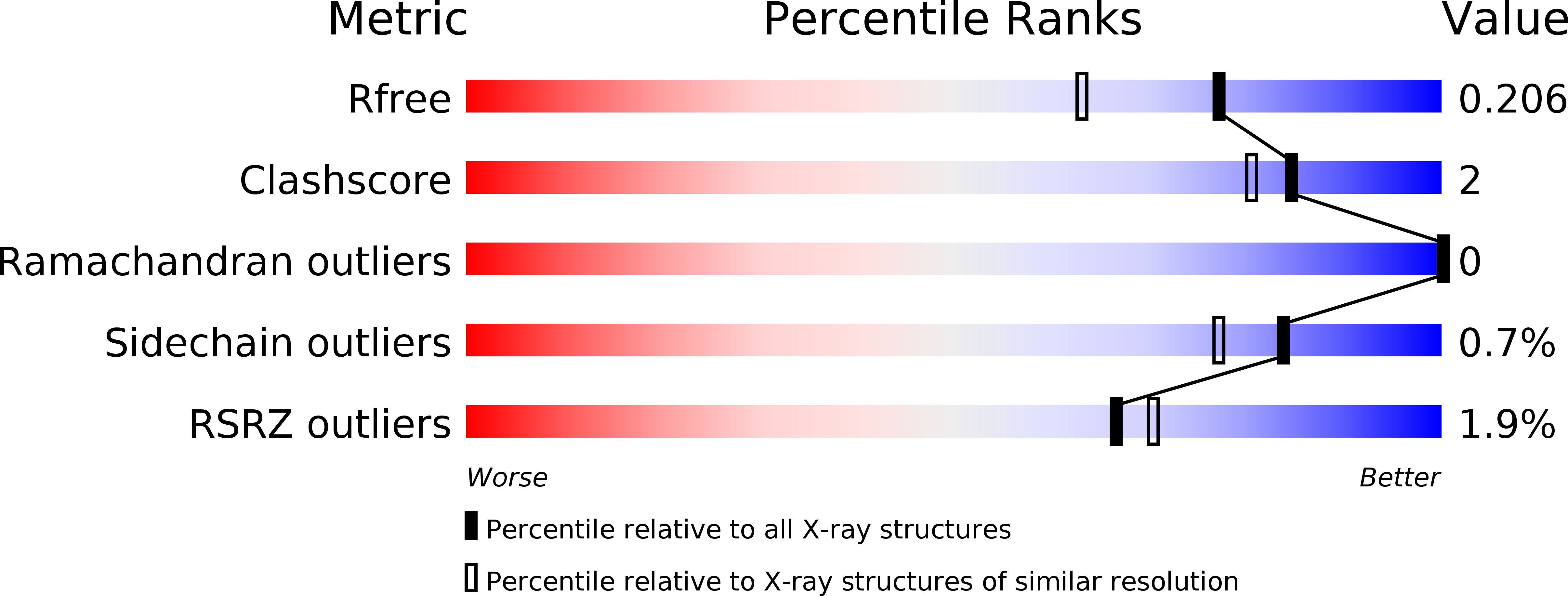
5YYA image
Entry Detail
PDB ID:
5YYA
Keywords:
Title:
Crystal structure of Tandem Tudor Domain of human UHRF1
Biological Source:
Source Organism:
Host Organism:
PDB Version:
Deposition Date:
2017-12-08
Release Date:
2018-12-12
Method Details:
Experimental Method:
Resolution:
1.70 Å
R-Value Free:
0.20
R-Value Work:
0.17
R-Value Observed:
0.17
Space Group:
P 21 21 21
Macromolecular Entities
Polymer Type:polypeptide(L)
Description:E3 ubiquitin-protein ligase UHRF1
Chain IDs:A
Chain Length:164
Number of Molecules:1
Biological Source:Homo sapiens
Primary Citation
Structure of the UHRF1 Tandem Tudor Domain Bound to a Methylated Non-histone Protein, LIG1, Reveals Rules for Binding and Regulation.
Structure 27 485 ? (2019)
PMID: 30639225 DOI: 10.1016/j.str.2018.11.012

Abstact

The protein UHRF1 is crucial for DNA methylation maintenance. The tandem Tudor domain (TTD) of UHRF1 binds histone H3K9me2/3 with micromolar affinity, as well as unmethylated linker regions within UHRF1 itself, causing auto-inhibition. Recently, we showed that a methylated histone-like region of DNA ligase 1 (LIG1K126me2/me3) binds the UHRF1 TTD with nanomolar affinity, permitting UHRF1 recruitment to chromatin. Here we report the crystal structure of the UHRF1 TTD bound to a LIG1K126me3 peptide. The data explain the basis for the high TTD-binding affinity of LIG1K126me3 and reveal that the interaction may be regulated by phosphorylation. Binding of LIG1K126me3 switches the overall structure of UHRF1 from a closed to a flexible conformation, suggesting that auto-inhibition is relieved. Our results provide structural insight into how UHRF1 performs its key function in epigenetic maintenance.

Legend

Protein

Chemical

Disease

Primary Citation of related structures