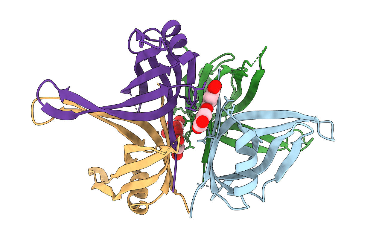
5YUN image
Entry Detail
PDB ID:
5YUN
Title:
Crystal structure of SSB complexed with myc
Biological Source:
PDB Version:
Deposition Date:
2017-11-22
Release Date:
2018-10-10
Method Details:
Experimental Method:
Resolution:
2.67 Å
R-Value Free:
0.25
R-Value Work:
0.20
R-Value Observed:
0.20
Space Group:
P 31
Macromolecular Entities
Polymer Type:polypeptide(L)
Description:Single-stranded DNA-binding protein
Chain IDs:A, B, C, D
Chain Length:121
Number of Molecules:4
Biological Source:Pseudomonas aeruginosa (strain ATCC 15692 / DSM 22644 / CIP 104116 / JCM 14847 / LMG 12228 / 1C / PRS 101 / PAO1)
Ligand Molecules
Primary Citation
Crystal structure of SSB complexed with inhibitor myricetin.
Biochem. Biophys. Res. Commun. 504 704 708 (2018)
PMID: 30213633 DOI: 10.1016/j.bbrc.2018.08.188

Abstact

Single-stranded DNA-binding protein (SSB) is essential for all DNA-dependent cellular processes. SSB inhibitors have been recently suggested as broad-spectrum antibacterial agents in antibiotic development. In this paper, we report the first inhibitor-complexed crystal structure of SSB from Pseudomonas aeruginosa PAO1 (PaSSB) at 2.68 Å resolution (PDB entry 5YUN). The inhibitor, myricetin, is a flavonol that possesses many pharmacological activities, such as anticancer, anti-inflammatory, and antibacterial properties, and is beneficial for humans. Four monomers of PaSSB and two of myricetins were found per asymmetric unit. Various interactions between myricetin and PaSSB were examined. Among these, four residues in PaSSB, Lys7, Arg62, Glu80, and Gly107 were found crucial for forming hydrogen bond to myricetin. These two myricetins occupy the grooves for ssDNA-binding of SSB that may prevent ssDNA-wrapping and ssDNA-binding stably from SSB. In addition to explaining how SSB can be inhibited, the myricetin-SSB interaction modes in this paper may also provide insights into how myricetin can bind and inhibit proteins on cancer-signaling pathways.

Legend

Protein

Chemical

Disease

Primary Citation of related structures