5YRZ image
Deposition Date 2017-11-11
Release Date 2018-05-23
Last Version Date 2024-03-27
Entry Detail
PDB ID:
5YRZ
Title:
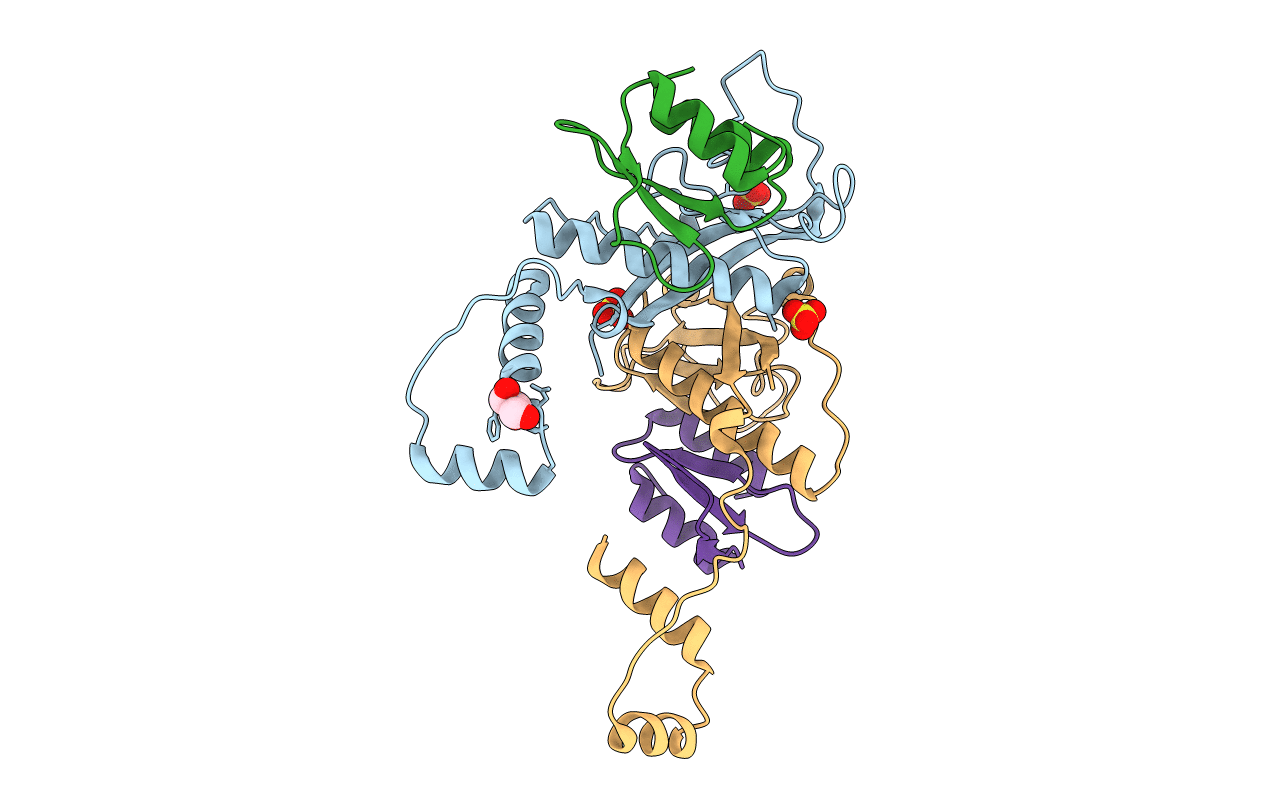
Toxin-Antitoxin complex from Streptococcus pneumoniae
Biological Source:
Method Details:
Experimental Method:
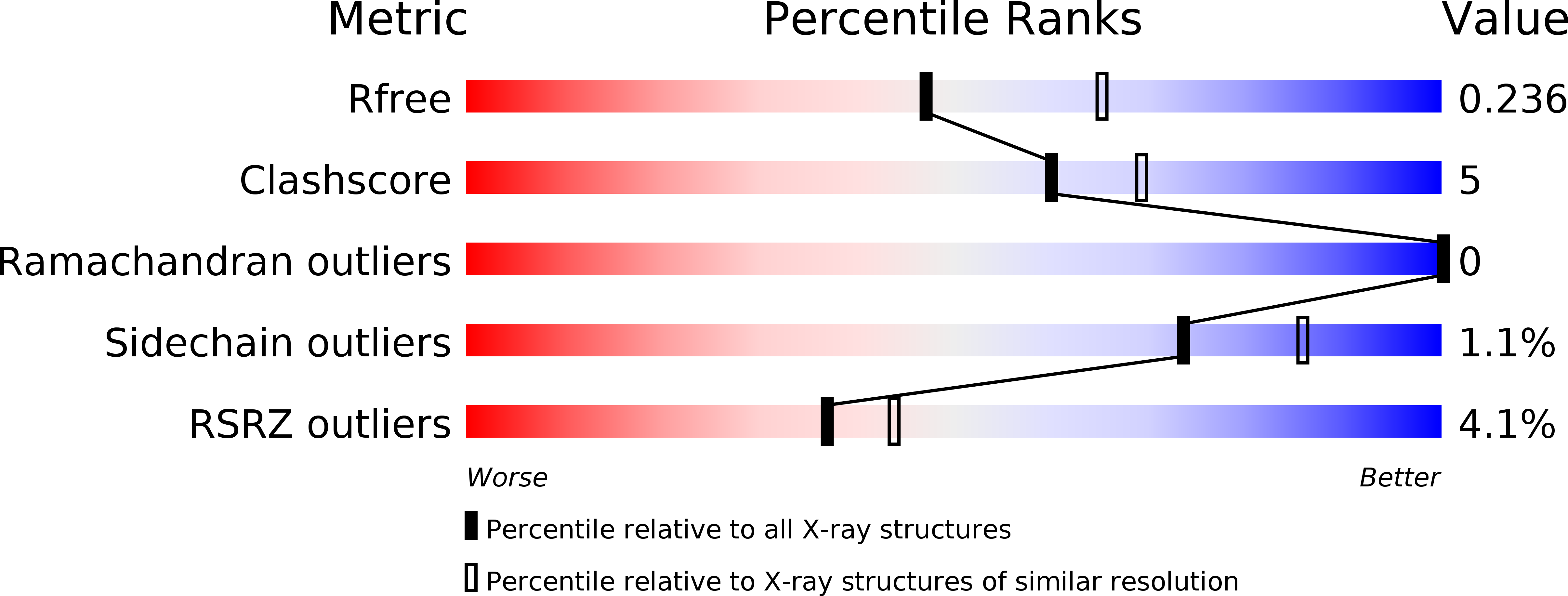
Resolution:
2.30 Å
R-Value Free:
0.23
R-Value Work:
0.20
R-Value Observed:
0.20
Space Group:
P 21 21 2
Macromolecular Entities
Polymer Type:polypeptide(L)
Molecule:HicB
Gene (Uniprot):SP_1786
Chain IDs:A, C
Chain Length:152
Number of Molecules:2
Biological Source:Streptococcus pneumoniae serotype 4 (strain ATCC BAA-334 / TIGR4)
Polymer Type:polypeptide(L)
Molecule:HicA
Gene (Uniprot):SP_1787
Chain IDs:B, D
Chain Length:89
Number of Molecules:2
Biological Source:Streptococcus pneumoniae serotype 4 (strain ATCC BAA-334 / TIGR4)
Primary Citation
Functional insights into the Streptococcus pneumoniae HicBA toxin-antitoxin system based on a structural study.
Nucleic Acids Res. 46 6371 6386 (2018)
PMID: 29878152 DOI: 10.1093/nar/gky469

Abstact

Streptococcus pneumonia has attracted increasing attention due to its resistance to existing antibiotics. TA systems are essential for bacterial persistence under stressful conditions such as nutrient deprivation, antibiotic treatment, and immune system attacks. In particular, S. pneumoniae expresses the HicBA TA gene, which encodes the stable HicA toxin and the labile HicB antitoxin. These proteins interact to form a non-toxic TA complex under normal conditions, but the toxin is activated by release from the antitoxin in response to unfavorable growth conditions. Here, we present the first crystal structure showing the complete conformation of the HicBA complex from S. pneumonia. The structure reveals that the HicA toxin contains a double-stranded RNA-binding domain that is essential for RNA recognition and that the C-terminus of the HicB antitoxin folds into a ribbon-helix-helix DNA-binding motif. The active site of HicA is sterically blocked by the N-terminal region of HicB. RNase activity assays show that His36 is essential for the ribonuclease activity of HicA, and nuclear magnetic resonance (NMR) spectra show that several residues of HicB participate in binding to the promoter DNA of the HicBA operon. A toxin-mimicking peptide that inhibits TA complex formation and thereby increases toxin activity was designed, providing a novel approach to the development of new antibiotics.

Legend

Protein

Chemical

Disease

Primary Citation of related structures