
Deposition Date
2017-11-10
Release Date
2018-03-07
Last Version Date
2024-11-13
Entry Detail
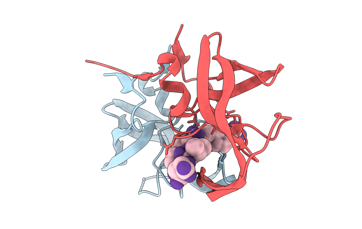
PDB ID:
5YRS
Keywords:
Title:
X-ray Snapshot of HIV-1 Protease in Action: Observation of Tetrahedral Intermediate and Its SIHB with Catalytic Aspartate
Biological Source:
Source Organism(s):
Human immunodeficiency virus 1 (Taxon ID: 11706)
Expression System(s):
Method Details:
Experimental Method:
Resolution:
1.76 Å
R-Value Free:
0.25
R-Value Work:
0.22
R-Value Observed:
0.22
Space Group:
P 61
