5YJ9 image
Deposition Date 2017-10-09
Release Date 2018-07-25
Last Version Date 2024-10-23
Entry Detail
PDB ID:
5YJ9
Keywords:
Title:
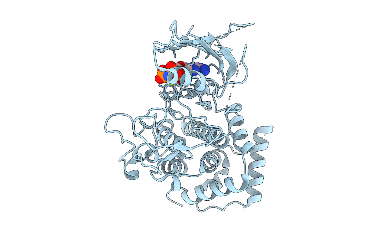
Crystal structure of Tribolium castaneum PINK1 kinase domain in complex with AMP-PNP
Biological Source:
Source Organism:
Host Organism:
Method Details:
Experimental Method:
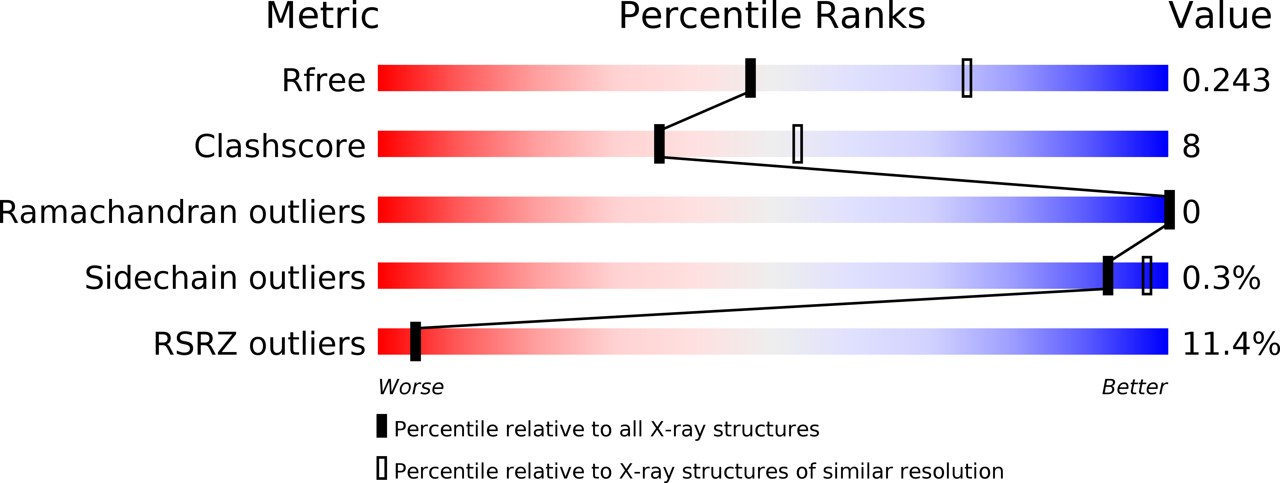
Resolution:
2.53 Å
R-Value Free:
0.24
R-Value Work:
0.22
R-Value Observed:
0.22
Space Group:
C 1 2 1
Macromolecular Entities
Structures with similar UniProt ID
Protein Blast
Polymer Type:polypeptide(L)
Molecule:Serine/threonine-protein kinase PINK1, mitochondrial-like Protein
Gene (Uniprot):Pink1
Mutagens:S205D, S377D, T386E, T530E
Chain IDs:A (auth: D)
Chain Length:423
Number of Molecules:1
Biological Source:Tribolium castaneum
Modified Residue
Compound ID Chain ID Parent Comp ID Details 2D Image
MSE A MET modified residue
Primary Citation
Structural insights into ubiquitin phosphorylation by PINK1.
Sci Rep 8 10382 10382 (2018)
PMID: 29991771 DOI: 10.1038/s41598-018-28656-8

Abstact

Mutations of PTEN-induced putative kinase 1 (PINK1) and the E3 ubiquitin (Ub) ligase parkin can cause familial parkinsonism. These two proteins are essential for ubiquitylation of damaged mitochondria and subsequent degradation. PINK1 phosphorylates Ser65 of Ub and the Ub-like (UBL) domain of parkin to allosterically relieve the autoinhibition of parkin. To understand the structural mechanism of the Ub/UBL-specific phosphorylation by PINK1, we determined the crystal structure of Tribolium castaneum PINK1 kinase domain (TcPINK1) in complex with a nonhydrolyzable ATP analogue at 2.5 Å resolution. TcPINK1 consists of the N- and C-terminal lobes with the PINK1-specific extension. The ATP analogue is bound in the cleft between the N- and C-terminal lobes. The adenine ring of the ATP analogue is bound to a hydrophobic pocket, whereas the triphosphate group of the ATP analogue and two coordinated Mg ions interact with the catalytic hydrophilic residues. Comparison with protein kinases A and C (PKA and PKC, respectively) unveils a putative Ub/UBL-binding groove, which is wider than the peptide-binding groove of PKA or PKC to accommodate the globular head of Ub or UBL. Further crosslinking analyses suggested a PINK1-interacting surface of Ub. Structure-guided mutational analyses support the findings from the present structural analysis of PINK1.

Legend

Protein

Chemical

Disease

Primary Citation of related structures
Feedback Form
Name
Email
Institute
Feedback