Deposition Date
2017-09-27
Release Date
2017-10-11
Last Version Date
2023-11-22
Entry Detail
PDB ID:
5YH7
Keywords:
Title:
Crystal structure of the complex of Phosphopantetheine adenylyltransferase from Acinetobacter baumannii with Coenzyme A at 2.0 A resolution
Biological Source:
Source Organism(s):
Acinetobacter baumannii (strain ACICU) (Taxon ID: 405416)
Expression System(s):
Method Details:
Experimental Method:
Resolution:
2.03 Å
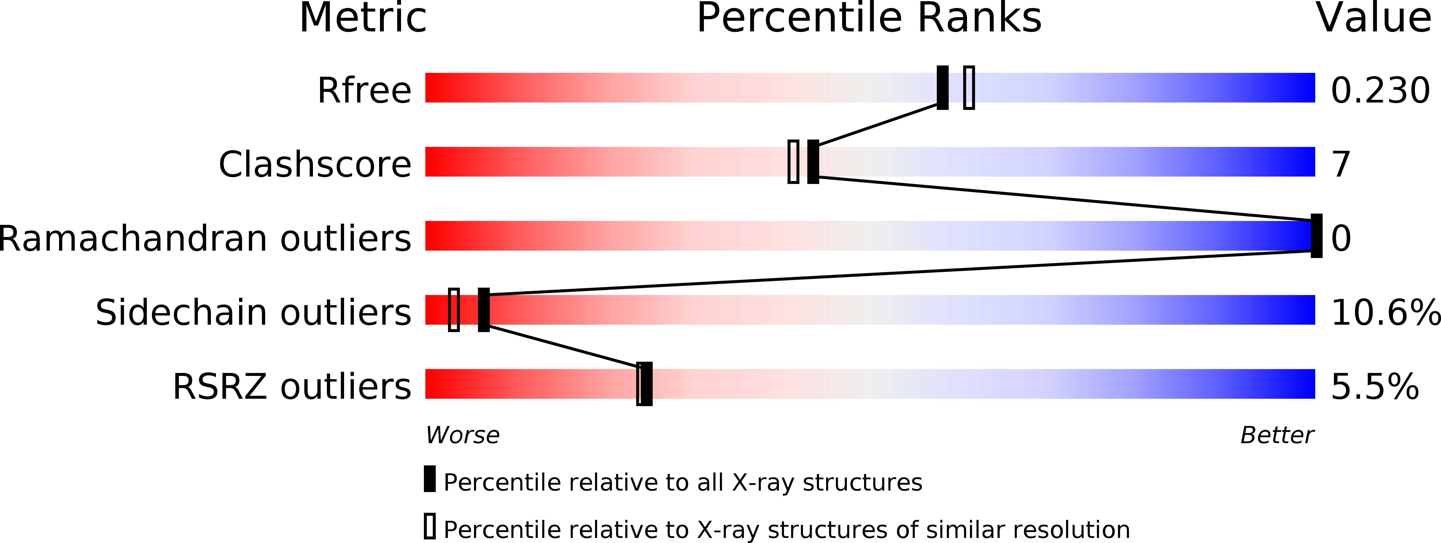
R-Value Free:
0.21
R-Value Work:
0.19
R-Value Observed:
0.19
Space Group:
F 41 3 2