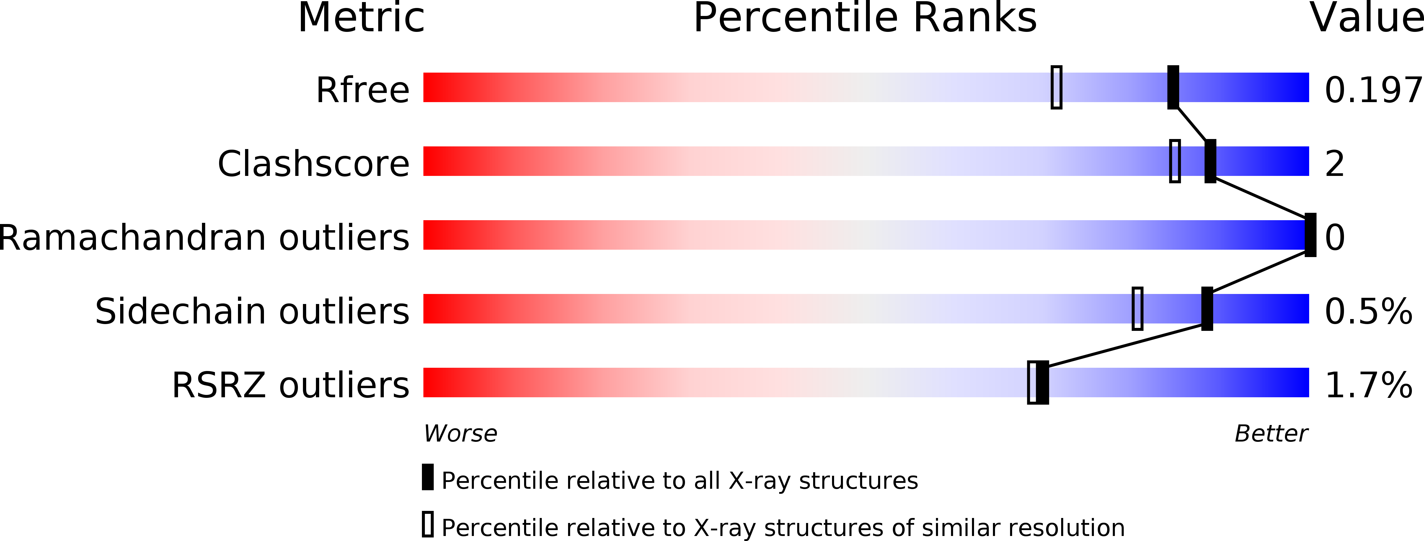
5YGM image
Deposition Date 2017-09-24
Release Date 2017-10-25
Last Version Date 2023-11-22
Entry Detail
PDB ID:
5YGM
Title:
Monomeric structure of concanavalin A at pH 7.5 from Carnivalia ensiformis
Biological Source:
Source Organism:
Host Organism:
Method Details:
Experimental Method:
Resolution:
1.60 Å
R-Value Free:
0.19
R-Value Work:
0.16
R-Value Observed:
0.16
Space Group:
I 2 2 2
Macromolecular Entities
Structures with similar UniProt ID
Protein Blast
Polymer Type:polypeptide(L)
Molecule:Concanavalin-A
Chain IDs:A
Chain Length:237
Number of Molecules:1
Biological Source:Canavalia ensiformis
Primary Citation
Heterometal-Coordinated Monomeric Concanavalin A at pH 7.5 from Canavalia ensiformis
J. Microbiol. Biotechnol. 27 2241 2244 (2017)
PMID: 29025256 DOI: 10.4014/jmb.1709.09057

Abstact

The structure of concanavalin A (ConA) has been studied intensively owing to its specific interactions with carbohydrates and its heterometal (Ca²⁺ and Mn²⁺) coordination. Most structures from X-ray crystallography have shown ConA as a dimer or tetramer, because the complex formation requires specific crystallization conditions. Here, we reported the monomeric structure of ConA with a resolution of 1.6 Å, which revealed that metal coordination could trigger sugar-binding ability. The calcium coordination residue, Asn14, changed the orientation of carbohydrate-binding residues and biophysical details, including structural information, providing valuable clues for the development and application of detection kits using ConA.

Legend

Protein

Chemical

Disease

Primary Citation of related structures