5YBB image
Deposition Date 2017-09-04
Release Date 2017-11-29
Last Version Date 2024-11-06
Entry Detail
PDB ID:
5YBB
Title:
Structural basis underlying complex assembly andconformational transition of the type I R-M system
Biological Source:
Method Details:
Experimental Method:
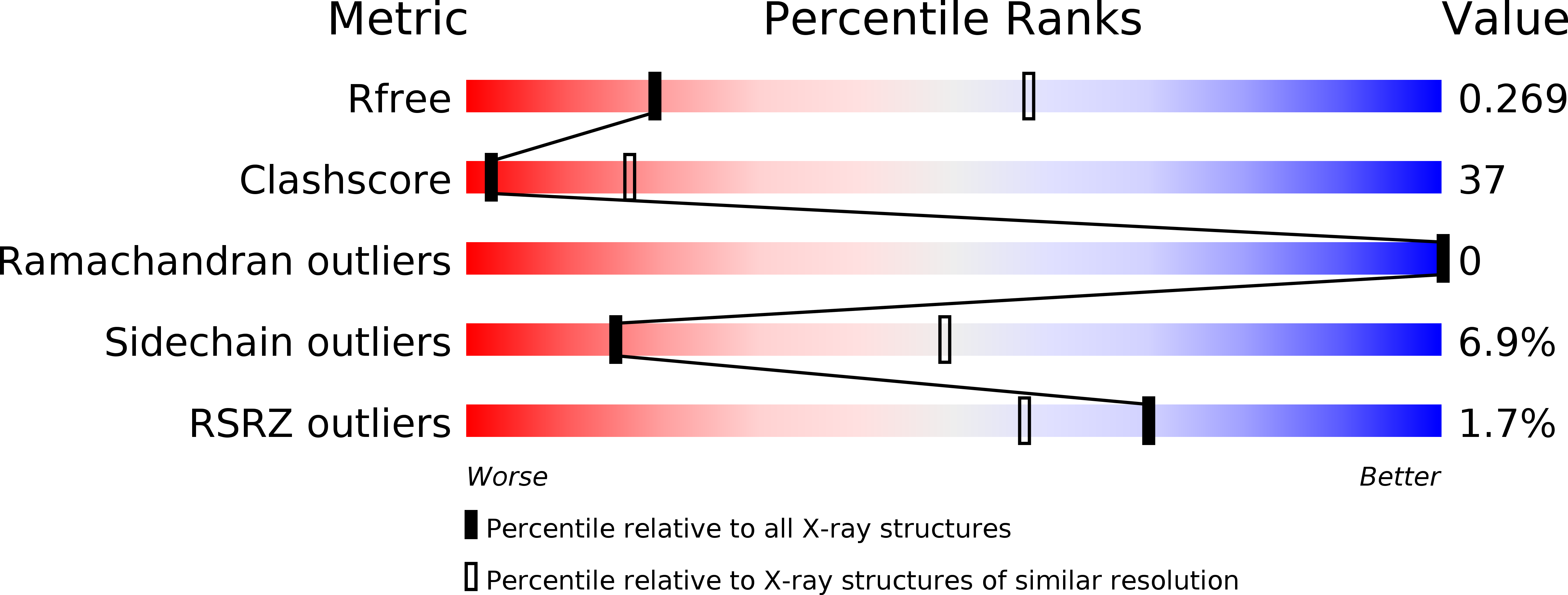
Resolution:
3.20 Å
R-Value Free:
0.27
R-Value Work:
0.23
R-Value Observed:
0.23
Space Group:
P 41
Macromolecular Entities
Structures with similar UniProt ID
Protein Blast
Polymer Type:polypeptide(L)
Molecule:Type I restriction-modification system methyltransferase subunit
Gene (Uniprot):HsdM
Chain IDs:A, B (auth: C), D (auth: B), E
Chain Length:507
Number of Molecules:4
Biological Source:Caldanaerobacter subterraneus subsp. tengcongensis (strain DSM 15242 / JCM 11007 / NBRC 100824 / MB4)
Structures with similar UniProt ID
Protein Blast
Polymer Type:polypeptide(L)
Molecule:Restriction endonuclease S subunits
Gene (Uniprot):HsdS
Chain IDs:C (auth: D), F (auth: G)
Chain Length:398
Number of Molecules:2
Biological Source:Caldanaerobacter subterraneus subsp. tengcongensis
Polymer Type:polydeoxyribonucleotide
Molecule:DNA
Chain IDs:G (auth: H)
Chain Length:22
Number of Molecules:1
Biological Source:synthetic construct
Polymer Type:polydeoxyribonucleotide
Molecule:DNA
Chain IDs:H (auth: I)
Chain Length:22
Number of Molecules:1
Biological Source:synthetic construct
Ligand Molecules
Primary Citation
Structural basis underlying complex assembly and conformational transition of the type I R-M system.
Proc. Natl. Acad. Sci. U.S.A. 114 11151 11156 (2017)
PMID: 28973912 DOI: 10.1073/pnas.1711754114

Abstact

Type I restriction-modification (R-M) systems are multisubunit enzymes with separate DNA-recognition (S), methylation (M), and restriction (R) subunits. Despite extensive studies spanning five decades, the detailed molecular mechanisms underlying subunit assembly and conformational transition are still unclear due to the lack of high-resolution structural information. Here, we report the atomic structure of a type I MTase complex (2M+1S) bound to DNA and cofactor S-adenosyl methionine in the "open" form. The intermolecular interactions between M and S subunits are mediated by a four-helix bundle motif, which also determines the specificity of the interaction. Structural comparison between open and previously reported low-resolution "closed" structures identifies the huge conformational changes within the MTase complex. Furthermore, biochemical results show that R subunits prefer to load onto the closed form MTase. Based on our results, we proposed an updated model for the complex assembly. The work reported here provides guidelines for future applications in molecular biology.

Legend

Protein

Chemical

Disease

Primary Citation of related structures