5Y97 image
Entry Detail
PDB ID:
5Y97
Keywords:
Title:
Crystal structure of snake gourd seed lectin in complex with lactose
Biological Source:
Source Organism:
PDB Version:
Deposition Date:
2017-08-23
Release Date:
2018-08-29
Method Details:
Experimental Method:
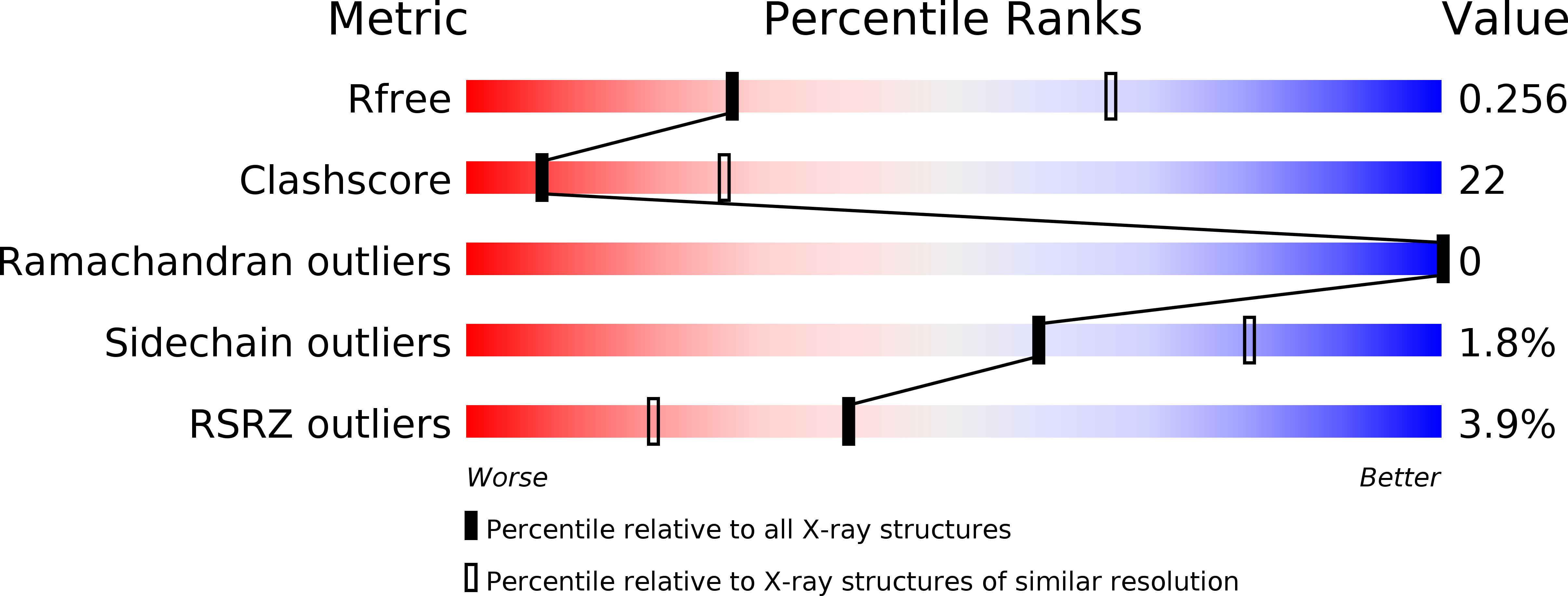
Resolution:
3.05 Å
R-Value Free:
0.25
R-Value Work:
0.22
R-Value Observed:
0.22
Space Group:
P 61 2 2
Macromolecular Entities
Polymer Type:polypeptide(L)
Description:Seed lectin
Chain IDs:A
Chain Length:41
Number of Molecules:1
Biological Source:Trichosanthes anguina
Polymer Type:polypeptide(L)
Description:Seed lectin
Chain IDs:B
Chain Length:209
Number of Molecules:1
Biological Source:Trichosanthes anguina
Polymer Type:polypeptide(L)
Description:Seed lectin
Chain IDs:C
Chain Length:264
Number of Molecules:1
Biological Source:Trichosanthes anguina
Ligand Molecules
Peptide-like Molecules
PRD_900004
Primary Citation
Ligand binding and retention in snake gourd seed lectin (SGSL). A crystallographic, thermodynamic and molecular dynamics study
Glycobiology 28 968 977 (2018)
PMID: 30099481 DOI: 10.1093/glycob/cwy072

Abstact

Snake gourd seed lectin (SGSL) is a non-toxic homolog of type II ribosome-inactivating proteins (RIPs) which contain a catalytic domain and a lectin domain. Isothermal titration calorimetry (ITC) measurements of the interactions of the protein with LacNAc, Lac, Gal, Me-α-Gal were carried out and the crystal structures of the native protein and its complex with Lac were determined. The crystal structure of the Me-α-Gal complex has already been determined. While the crystal structure showed the presence of two-sugar-binding sites, one on each of the two domains of the lectin chain, ITC measurements indicated the presence of only one binding site. In order to resolve this anomaly, molecular dynamics (MD) simulations were carried out on the native protein and on its complexes with Me-α-Gal and Lac. Simulations were also performed on the protein after reducing the inter-chain disulfide bridge between the two chains. The crystal structures and the simulations confirmed the robustness of the protein structure, irrespective of the presence or absence of the disulfide bridge. The simulations indicated that although two sites can bind sugar, only the ligand at one site is retained in a dynamic situation. The studies thus bring out the subtle relationship between binding and retention of the ligand.

Legend

Protein

Chemical

Disease

Primary Citation of related structures