5Y7Z image
Deposition Date 2017-08-18
Release Date 2018-08-29
Last Version Date 2024-11-13
Entry Detail
PDB ID:
5Y7Z
Title:
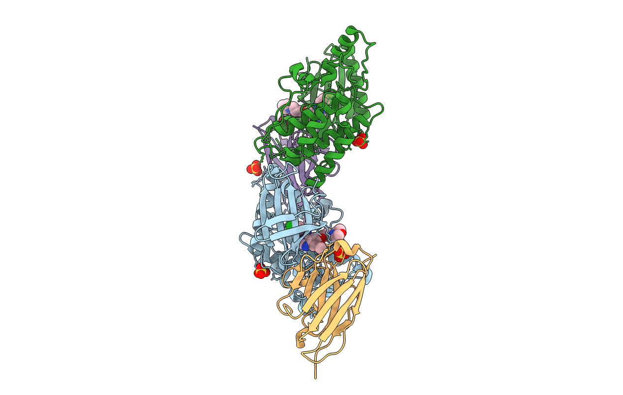
Complex structure of cyclin G-associated kinase with gefitinib
Biological Source:
Source Organism:
Homo sapiens (Taxon ID: 9606)
Lama glama (Taxon ID: 9844)
Host Organism:
Method Details:
Experimental Method:
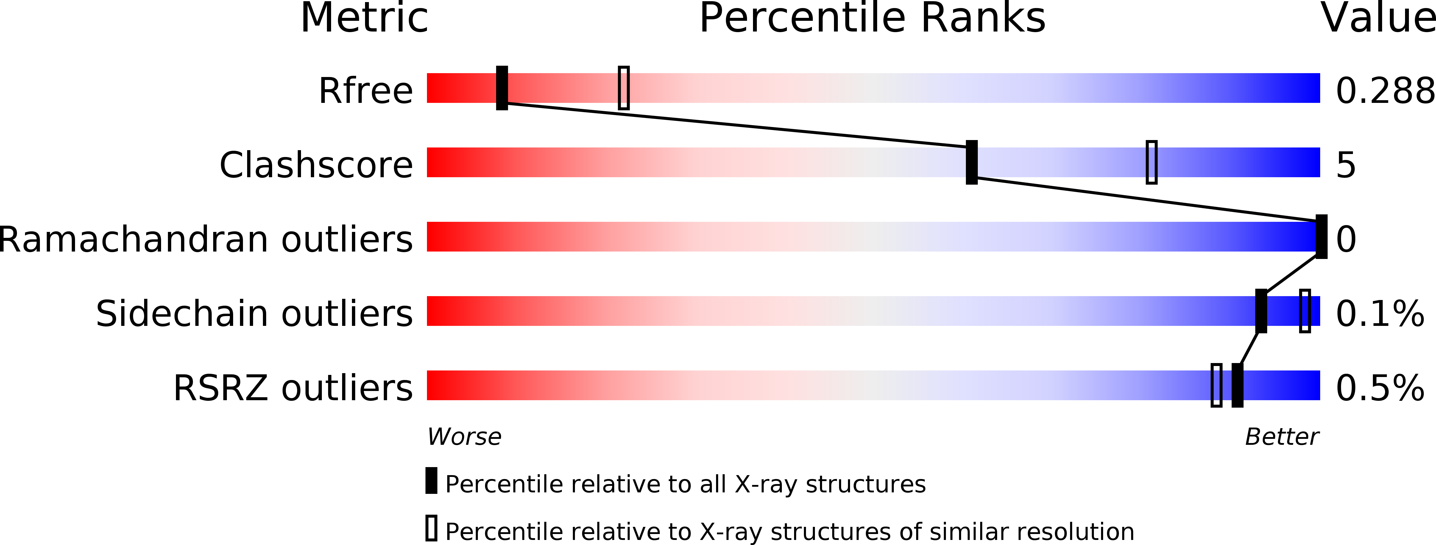
Resolution:
2.80 Å
R-Value Free:
0.28
R-Value Work:
0.23
R-Value Observed:
0.23
Space Group:
P 21 21 21
Macromolecular Entities
Polymer Type:polypeptide(L)
Molecule:Cyclin-G-associated kinase
Gene (Uniprot):GAK
Chain IDs:A, B
Chain Length:313
Number of Molecules:2
Biological Source:Homo sapiens
Polymer Type:polypeptide(L)
Molecule:NANOBODY
Chain IDs:C (auth: D), D (auth: C)
Chain Length:148
Number of Molecules:2
Biological Source:Lama glama
Primary Citation
Structural Basis for the Inhibition of Cyclin G-Associated Kinase by Gefitinib.
ChemistryOpen 7 721 727 (2018)
PMID: 30214852 DOI: 10.1002/open.201800177

Abstact

Gefitinib is the molecular target drug for advanced non-small-cell lung cancer. The primary target of gefitinib is the positive mutation of epidermal growth factor receptor, but it also inhibits cyclin G-associated kinase (GAK). To reveal the molecular bases of GAK and gefitinib binding, structure analyses were conducted and determined two forms of the gefitinib-bound nanobody⋅GAK kinase domain complex structures. The first form, GAK_1, has one gefitinib at the ATP binding pocket, whereas the second form, GAK_2, binds one each in the ATP binding site and a novel binding site adjacent to the activation segment C-terminal helix, a unique element of the Numb-associated kinase family. In the novel binding site, gefitinib binds in the hydrophobic groove around the activation segment, disrupting the conserved hydrogen bonds for the catalytic activity. These structures suggest possibilities for the development of selective GAK inhibitors for viral infections, such as the hepatitis C virus.

Legend

Protein

Chemical

Disease

Primary Citation of related structures