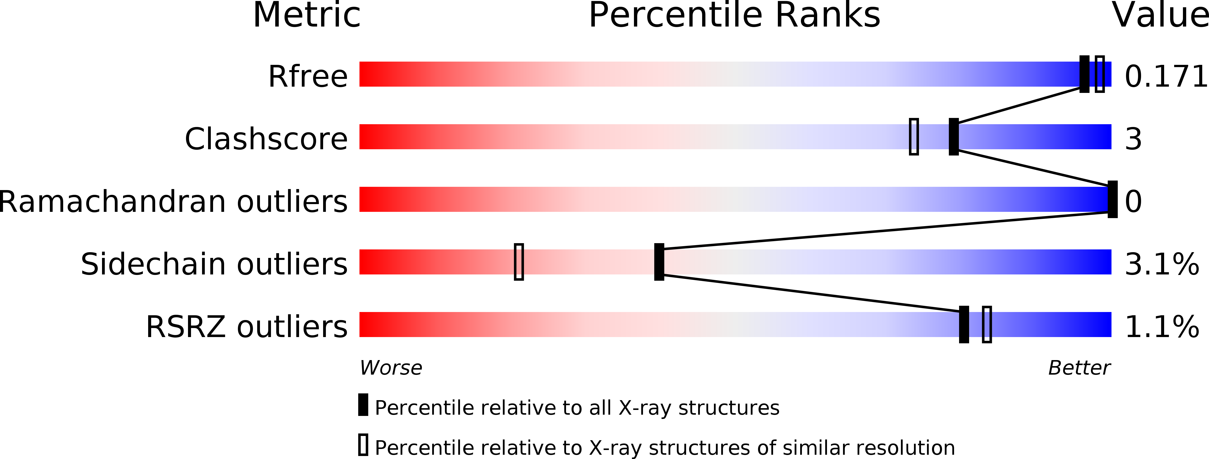
5Y6T image
Entry Detail
PDB ID:
5Y6T
Keywords:
Title:
Crystal structure of endo-1,4-beta-mannanase from Eisenia fetida
Biological Source:
Source Organism:
PDB Version:
Deposition Date:
2017-08-15
Release Date:
2018-06-27
Method Details:
Experimental Method:
Resolution:
1.70 Å
R-Value Free:
0.16
R-Value Work:
0.13
R-Value Observed:
0.13
Space Group:
P 21 21 21
Macromolecular Entities
Polymer Type:polypeptide(L)
Description:endo-1,4-beta-mannanase
Chain IDs:A
Chain Length:387
Number of Molecules:1
Biological Source:Eisenia fetida
Primary Citation
Gene cloning, expression, and X-ray crystallographic analysis of a beta-mannanase from Eisenia fetida.
Enzyme.Microb.Technol. 117 15 22 (2018)
PMID: 30037547 DOI: 10.1016/j.enzmictec.2018.05.014

Abstact

The endo-1,4-β-mannanases (Ef-Man) gene from Eisenia fetida was determined to consist of 1131 bp and encode a 377 amino acid protein. The amino acid sequence showed similarity with the endo-1,4-β-mannanases of Daphnia pulex (62%), Cryptopygus antarcticus (64%), Crassostrea gigas (61%), Mytilus edulis (60%), and Aplysia kurodai (58%). The gene encoding mature Ef-Man was expressed in Pichia pastoris (GS115 strain). Based on SDS-PAGE analysis, the molecular mass of the purified recombinant Ef-Man (rEf-Man) was estimated to be 39 kDa. All catalytically important residues of endo-1,4-β-mannanases in the glycoside hydrolase (GH) family 5 were conserved in Ef-Man. The optimal temperature for rEf-Man was identified as 60 °C. HPLC and HPAEC analyses suggest that Ef-Man requires at least six subsites for efficient hydrolysis and is capable of performing transglycosylation reactions. The overall structure of rEf-Man is similar to those of GH5 family proteins, and tertiary structures around the active site are conserved among endo-1,4-β-mannanase families. X-ray crystallographic analysis supports the hydrolysis and transglycosylation reaction mechanism determined by HPLC and HPAEC analyses.

Legend

Protein

Chemical

Disease

Primary Citation of related structures