5Y5W image
Deposition Date 2017-08-10
Release Date 2017-10-11
Last Version Date 2023-11-22
Entry Detail
PDB ID:
5Y5W
Keywords:
Title:
Crystal structure of human Spindlin1 in complex with a histone H4K20(me3) peptide
Biological Source:
Source Organism:
Homo sapiens (Taxon ID: 9606)
Host Organism:
Method Details:
Experimental Method:
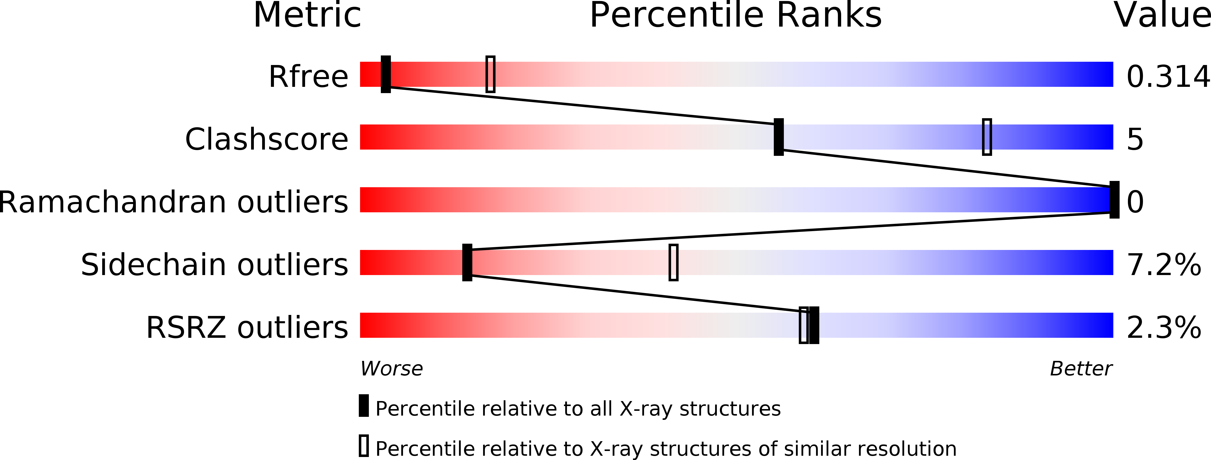
Resolution:
3.30 Å
R-Value Free:
0.30
R-Value Work:
0.21
R-Value Observed:
0.22
Space Group:
C 2 2 21
Macromolecular Entities
Structures with similar UniProt ID
Protein Blast
Polymer Type:polypeptide(L)
Molecule:Spindlin-1
Gene (Uniprot):SPIN1
Chain IDs:A, B, C, D
Chain Length:235
Number of Molecules:4
Biological Source:Homo sapiens
Polymer Type:polypeptide(L)
Molecule:Histone peptide H4K20(me3)
Chain IDs:E, F, G
Chain Length:9
Number of Molecules:3
Biological Source:Homo sapiens
Ligand Molecules
Primary Citation
Spindlin-1 recognizes methylations of K20 and R23 of histone H4 tail
FEBS Lett. 592 4098 4110 (2018)
PMID: 30381828 DOI: 10.1002/1873-3468.13281

Abstact

Using methods combining cross-linking, pull-down assays, and stable isotope labeling by amino acids in cell culture with mass spectrometry, we identified that the Tudor domain-containing protein Spindlin-1 recognizes trimethylation of histone H4 lysine 20 (H4K20me3). The binding affinity of Spindlin-1 to H4K20me3 is weaker than that to H3K4me3, indicating H4K20me3 as a secondary substrate for Spindlin-1. Structural studies of Spindlin-1 in complex with the H4K20me3 peptide indicate that Spindlin-1 attains a distinct binding mode for H4K20me3 recognition. Further biochemical analysis identified that Spindlin-1 also binds methylated R23 of H4, providing new clues for the function of Spindlin-1.

Legend

Protein

Chemical

Disease

Primary Citation of related structures
Feedback Form
Name
Email
Institute
Feedback