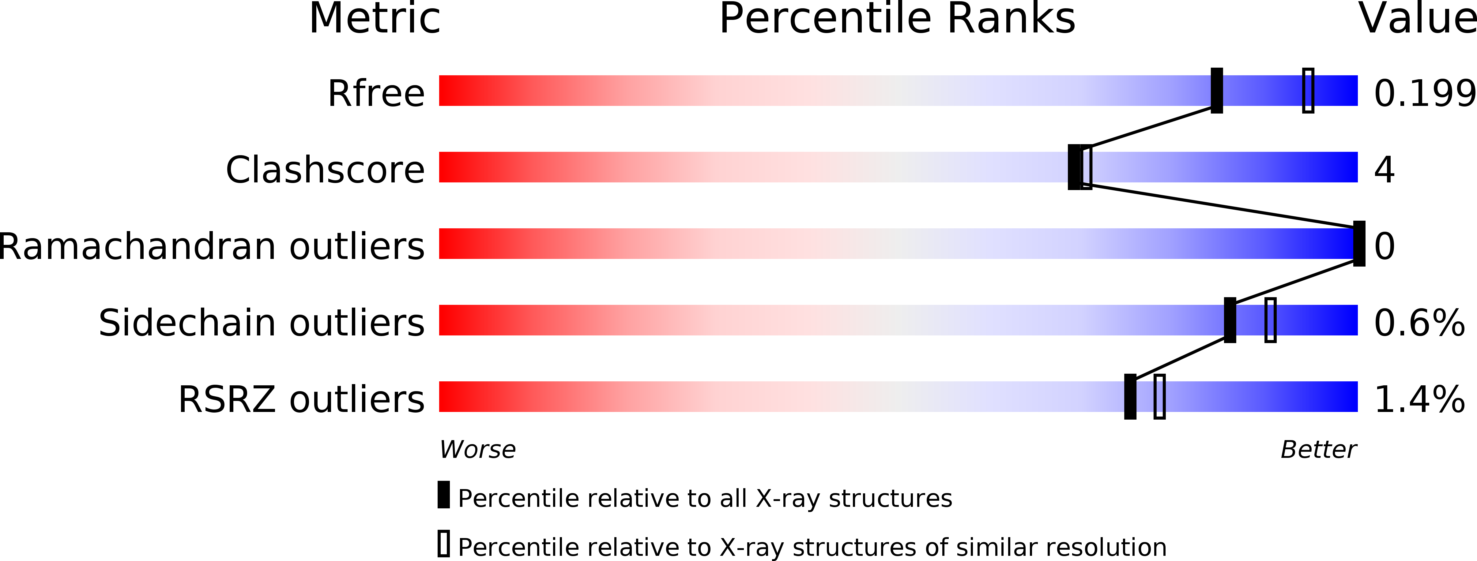
5Y5M image
Deposition Date 2017-08-09
Release Date 2018-08-08
Last Version Date 2023-11-22
Entry Detail
PDB ID:
5Y5M
Keywords:
Title:
SFX structure of cytochrome P450nor: a complete dark data without pump laser (resting state)
Biological Source:
Source Organism:
Host Organism:
Method Details:
Experimental Method:
Resolution:
2.10 Å
R-Value Free:
0.21
R-Value Work:
0.16
R-Value Observed:
0.16
Space Group:
P 1 21 1
Macromolecular Entities
Polymer Type:polypeptide(L)
Molecule:NADP nitrous oxide-forming nitric oxide reductase
Gene (Uniprot):CYP55A1
Chain IDs:A, B
Chain Length:403
Number of Molecules:2
Biological Source:Fusarium oxysporum
Ligand Molecules
Primary Citation
Capturing an initial intermediate during the P450nor enzymatic reaction using time-resolved XFEL crystallography and caged-substrate.
Nat Commun 8 1585 1585 (2017)
PMID: 29147002 DOI: 10.1038/s41467-017-01702-1

Abstact

Time-resolved serial femtosecond crystallography using an X-ray free electron laser (XFEL) in conjunction with a photosensitive caged-compound offers a crystallographic method to track enzymatic reactions. Here we demonstrate the application of this method using fungal NO reductase, a heme-containing enzyme, at room temperature. Twenty milliseconds after caged-NO photolysis, we identify a NO-bound form of the enzyme, which is an initial intermediate with a slightly bent Fe-N-O coordination geometry at a resolution of 2.1 Å. The NO geometry is compatible with those analyzed by XFEL-based cryo-crystallography and QM/MM calculations, indicating that we obtain an intact Fe3+-NO coordination structure that is free of X-ray radiation damage. The slightly bent NO geometry is appropriate to prevent immediate NO dissociation and thus accept H- from NADH. The combination of using XFEL and a caged-compound is a powerful tool for determining functional enzyme structures during catalytic reactions at the atomic level.

Legend

Protein

Chemical

Disease

Primary Citation of related structures