Deposition Date
2017-08-01
Release Date
2017-09-20
Last Version Date
2024-10-23
Entry Detail
PDB ID:
5Y48
Keywords:
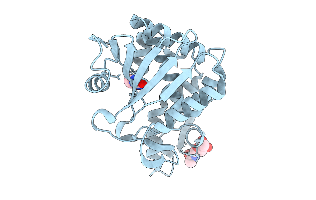
Title:
Crystal structure of the complex of Ribosome inactivating protein from Momordica balsamina with Pyrimidine-2,4-dione at 1.70 Angstrom resolution
Biological Source:
Source Organism(s):
Momordica balsamina (Taxon ID: 3672)
Method Details:
Experimental Method:
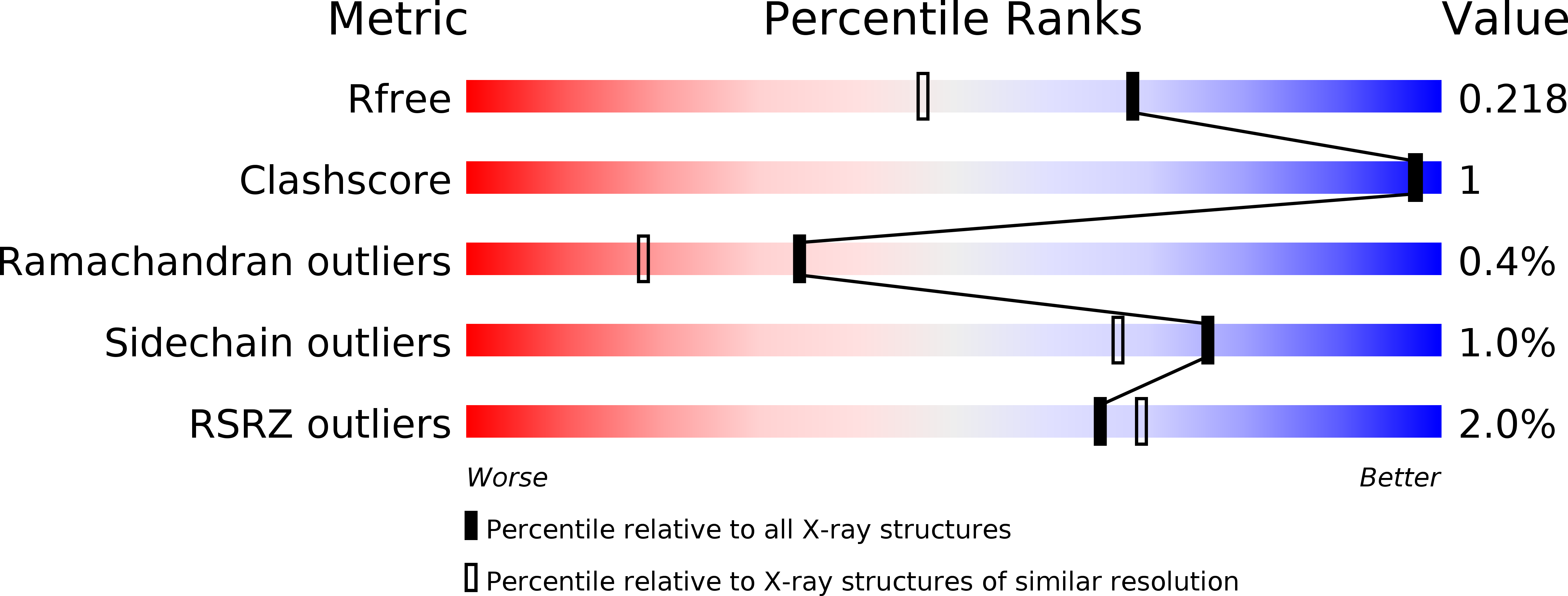
Resolution:
1.70 Å
R-Value Free:
0.21
R-Value Work:
0.16
R-Value Observed:
0.16
Space Group:
H 3