5Y40 image
Deposition Date 2017-07-31
Release Date 2018-04-11
Last Version Date 2023-11-22
Entry Detail
PDB ID:
5Y40
Keywords:
Title:
Structure of the periplasmic domain of the MotB L119P mutant from Salmonella (crystal form 2)
Biological Source:
Method Details:
Experimental Method:
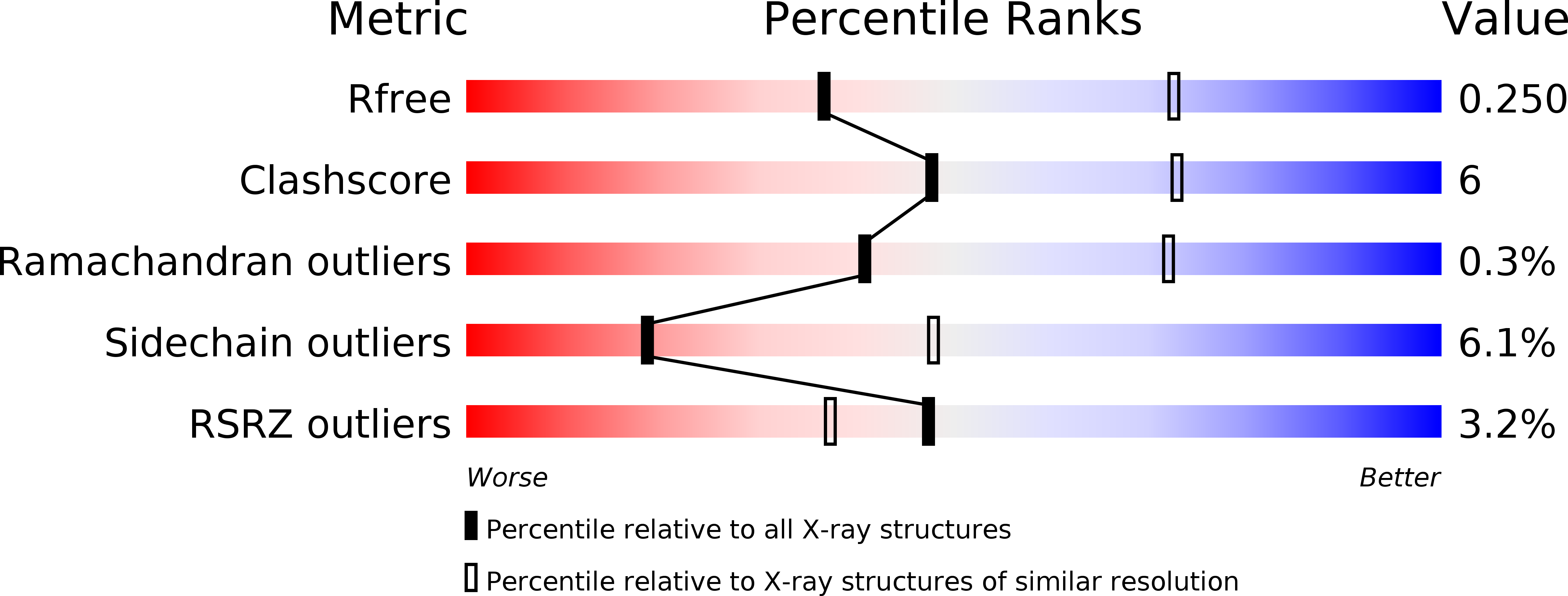
Resolution:
2.80 Å
R-Value Free:
0.25
R-Value Work:
0.21
R-Value Observed:
0.21
Space Group:
P 4 21 2
Macromolecular Entities
Polymer Type:polypeptide(L)
Molecule:Motility protein B
Gene (Uniprot):motB
Mutations:L119P
Chain IDs:A, B, C, D
Chain Length:183
Number of Molecules:4
Biological Source:Salmonella typhimurium (strain LT2 / SGSC1412 / ATCC 700720)
Ligand Molecules
Primary Citation
The Helix Rearrangement in the Periplasmic Domain of the Flagellar Stator B Subunit Activates Peptidoglycan Binding and Ion Influx.
Structure 26 590 598.e5 (2018)
PMID: 29576320 DOI: 10.1016/j.str.2018.02.016

Abstact

The stator of the bacterial flagellar motor couples ion flow with torque generation. The ion-conducting stator channel opens only when incorporated into and anchored around the rotor via the peptidoglycan (PG) binding domain of the B subunit (MotBC). However, no direct evidence of PG binding coupled with channel activation has been presented. Here, we report the structural rearrangements of MotBC responsible for this coupling process. A MotBC fragment with the L119P replacement, which is known to cause channel activation, was able to bind PG. Nuclear magnetic resonance analysis of MotBC and the crystal structure of the MotBC-L119P dimer revealed major structural changes in helix α1. In vivo crosslinking results confirm that a major rearrangement occurs. Our results suggest that, upon stator incorporation into the motor, helix α1 of MotBC changes into an extended non-helical structure. We propose that this change allows the stator both to bind PG and to open its proton channel.

Legend

Protein

Chemical

Disease

Primary Citation of related structures