5Y2E image
Deposition Date 2017-07-25
Release Date 2018-03-14
Last Version Date 2024-03-27
Entry Detail
PDB ID:
5Y2E
Keywords:
Title:
Crystal structure of the oligomerization domain of NSP4 from the rotavirus strain NCDV
Biological Source:
Method Details:
Experimental Method:
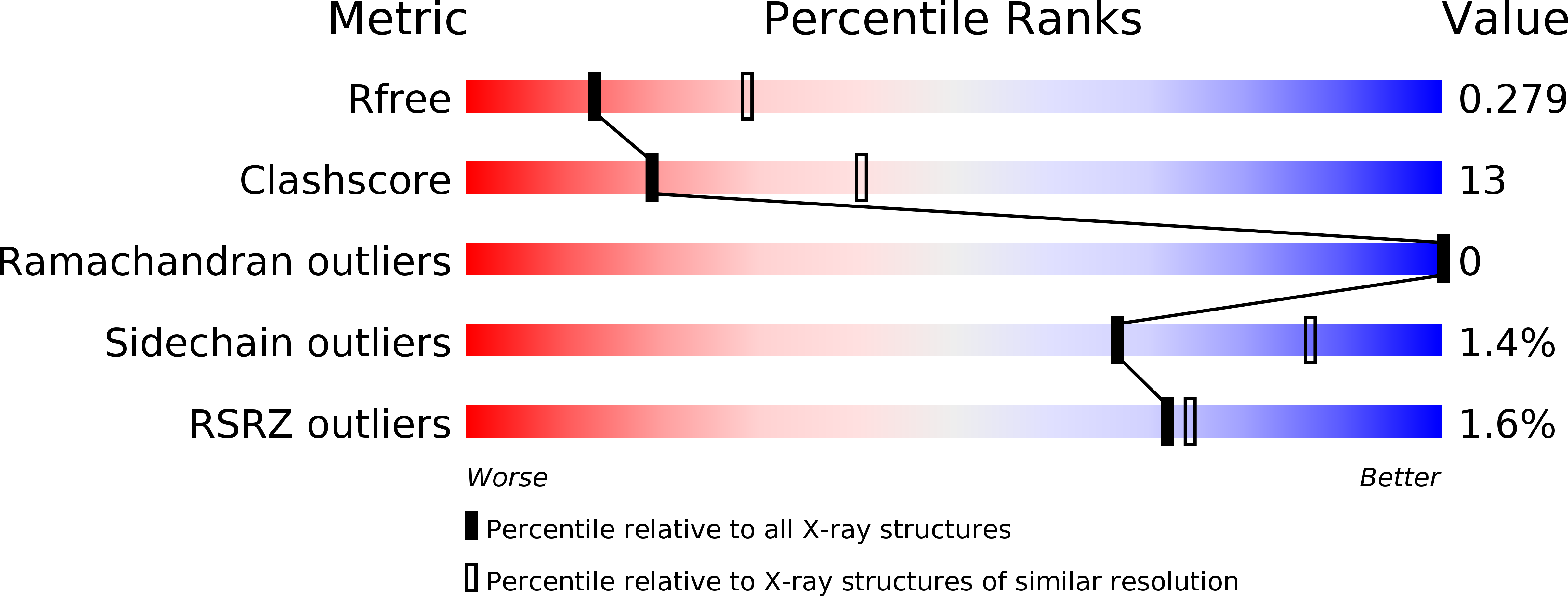
Resolution:
2.70 Å
R-Value Free:
0.27
R-Value Work:
0.23
R-Value Observed:
0.23
Space Group:
P 21 21 21
Macromolecular Entities
Structures with similar UniProt ID
Protein Blast
Polymer Type:polypeptide(L)
Molecule:Non-structural glycoprotein 4
Mutagens:E120G
Chain IDs:A (auth: D), B (auth: A), C, D (auth: B)
Chain Length:47
Number of Molecules:4
Biological Source:Rotavirus A (strain RVA/Cow/United States/NCDV-Lincoln/1969/G6P6[1])
Primary Citation
New tetrameric forms of the rotavirus NSP4 with antiparallel helices.
Arch. Virol. 163 1531 1547 (2018)
PMID: 29455326 DOI: 10.1007/s00705-018-3753-6

Abstact

Rotavirus nonstructural protein 4, the first viral enterotoxin to be identified, is a multidomain, multifunctional glycoprotein. Earlier, we reported a Ca(2+)-bound coiled-coil tetrameric structure of the diarrhea-inducing region of NSP4 from the rotavirus strains SA11 and I321 and a Ca(2+)-free pentameric structure from the rotavirus strain ST3, all with a parallel arrangement of alpha-helices. pH was found to determine the oligomeric state: a basic pH favoured a tetramer, whereas an acidic pH favoured a pentamer. Here, we report two novel forms of the coiled-coil region of NSP4 from the bovine rotavirus strains MF66 and NCDV. These crystallized at acidic pH, forming antiparallel coiled-coil tetrameric structures without any bound Ca(2+) ion. Structural and mutational studies of the coiled-coil regions of NSP4 revealed that the nature of the residue at position 131 (Tyr/His) plays an important role in the observed structural diversity.

Legend

Protein

Chemical

Disease

Primary Citation of related structures