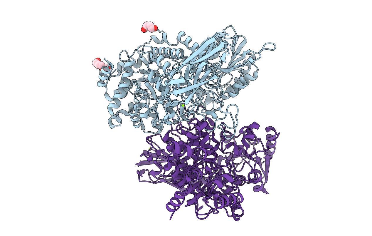
5XXL image
Deposition Date 2017-07-04
Release Date 2017-12-13
Last Version Date 2023-11-22
Entry Detail
PDB ID:
5XXL
Keywords:
Title:
Crystal structure of GH3 beta-glucosidase from Bacteroides thetaiotaomicron
Biological Source:
Method Details:
Experimental Method:
Resolution:
1.60 Å
R-Value Free:
0.22
R-Value Work:
0.20
R-Value Observed:
0.20
Space Group:
C 2 2 21
Macromolecular Entities
Polymer Type:polypeptide(L)
Molecule:Periplasmic beta-glucosidase
Gene (Uniprot):BT_3567
Chain IDs:A, B
Chain Length:760
Number of Molecules:2
Biological Source:Bacteroides thetaiotaomicron (strain ATCC 29148 / DSM 2079 / NCTC 10582 / E50 / VPI-5482)
Primary Citation
Function and structure relationships of a beta-1,2-glucooligosaccharide-degrading beta-glucosidase.
FEBS Lett. 591 3926 3936 (2017)
PMID: 29131329 DOI: 10.1002/1873-3468.12911

Abstact

BT_3567 protein, a putative β-glucosidase from Bacteroides thetaiotaomicron, exhibits higher activity toward Sop3-5 (Sopn , n: degree of polymerization of β-1,2-glucooligosaccharides) than toward Sop2 , unlike a known β-glucosidase from Listeria innocua which predominantly prefers Sop2 . In the complex structure determined by soaking of a D286N mutant crystal with Sop4 , a Sop3 moiety was observed at subsites -1 to +2. The glucose moiety at subsite +2 forms a hydrogen bond with Asn81, which is replaced with Gly in the L. innocua β-glucosidase. The Km values of the N81G mutant for Sop3-5 are much higher than those of the wild-type, suggesting that Asn81 contributes to the binding to substrates longer than Sop3 .

Legend

Protein

Chemical

Disease

Primary Citation of related structures