5XWB image
Deposition Date 2017-06-29
Release Date 2017-09-06
Last Version Date 2024-11-06
Entry Detail
PDB ID:
5XWB
Keywords:
Title:
Crystal Structure of 5-Enolpyruvulshikimate-3-phosphate Synthase from a Psychrophilic Bacterium, Colwellia psychrerythraea
Biological Source:
Method Details:
Experimental Method:
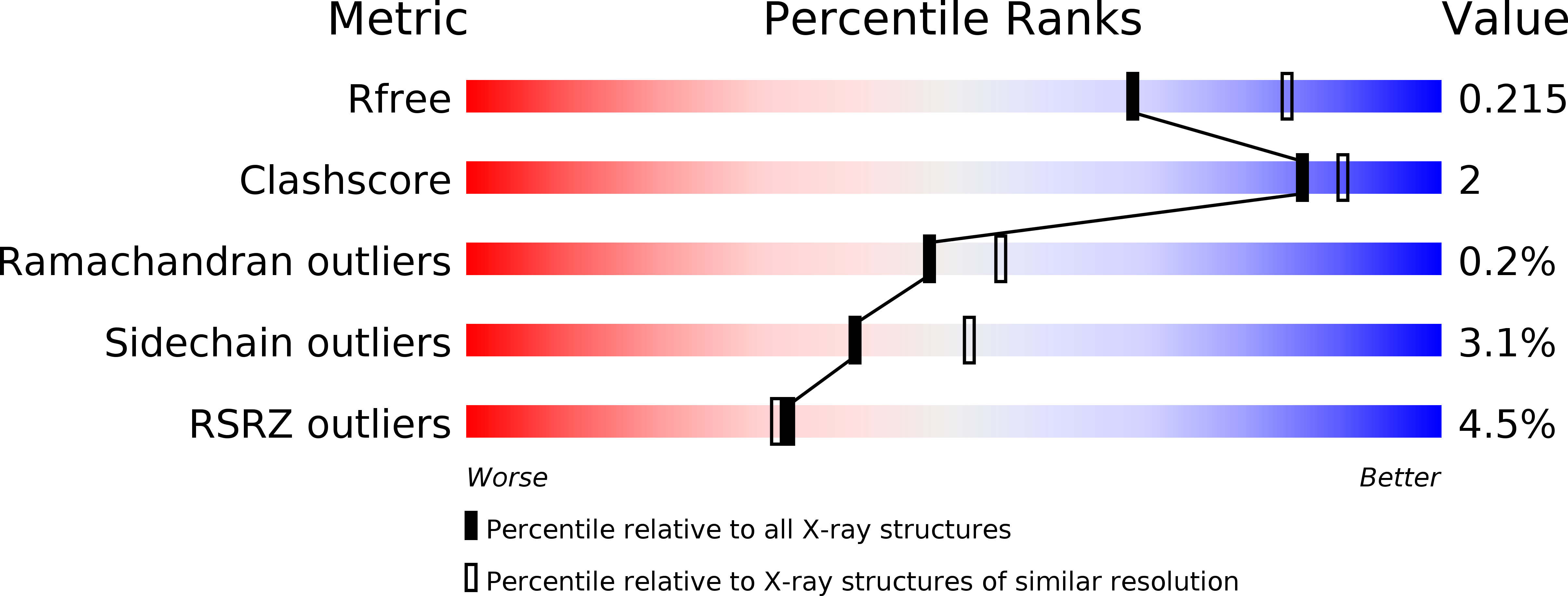
Resolution:
2.20 Å
R-Value Free:
0.20
R-Value Work:
0.14
R-Value Observed:
0.14
Space Group:
P 1 21 1
Macromolecular Entities
Structures with similar UniProt ID
Protein Blast
Polymer Type:polypeptide(L)
Molecule:3-phosphoshikimate 1-carboxyvinyltransferase
Gene (Uniprot):aroA
Chain IDs:A, B
Chain Length:446
Number of Molecules:2
Biological Source:Colwellia psychrerythraea (strain 34H / ATCC BAA-681)
Primary Citation
Crystal structure of 5-enolpyruvylshikimate-3-phosphate synthase from a psychrophilic bacterium, Colwellia psychrerythraea 34H.
Biochem. Biophys. Res. Commun. 492 500 506 (2017)
PMID: 28830812 DOI: 10.1016/j.bbrc.2017.08.063

Abstact

To survive at low temperatures, psychrophiles seem to produce cold-adapted enzymes with a high flexibility around active sites for high catalytic efficiency. To gain insights into the cold-adaptation of psychrophilic enzymes in atomic detail, we determined the crystal structure of 5-enolpyruvylshikimate-3-phosphate synthase (CpsEPSPS) from Colwellia psychrerythraea, a psychrophilic bacterium. EPSPS is the primary target for the broad-spectrum herbicide, glyphosate, and a promising target for the development of antimicrobial and antiparasitic agents since it is absent in animals. The crystal structure of unliganded, open CpsEPSPS was determined at 2.2 Å resolution in space group P21 with two protomers per asymmetric unit. Superposition of separate domain I and II of CpsEPSPS structure with those of Escherichia coli EPSPS (EcoEPSPS) structure showed relatively small differences of RMSD values of 0.423 Å and 0.693 Å for domains I and II, respectively, implying the residues in ligand binding and catalysis of cold-adapted CpsEPSPS showed no significant flexibility. This result is conflicting to other cases of cold-adapted proteins. We also observed that hydrogen-bond forming residues in the surface of EcoEPSPS was mutated to non- or lesser hydrogen-bond forming one in CpsEPSPS, which makes the protein surface softer and eventually makes the protein more active at low temperature. In addition, domain rotation angle between open and closed states of CpsEPSPS was smaller than those of any EPSPSs whose structures are known. The restriction of the domain closure, which reduces the entropy cost of ligand binding and catalysis, may be a novel molecular adaptations of cold-adapted enzymes.

Legend

Protein

Chemical

Disease

Primary Citation of related structures