5XTU image
Deposition Date 2017-06-21
Release Date 2018-10-10
Last Version Date 2025-04-09
Entry Detail
PDB ID:
5XTU
Keywords:
Title:
Crystal Structure of GDSL Esterase of Photobacterium sp. J15
Biological Source:
Source Organism:
Host Organism:
Method Details:
Experimental Method:
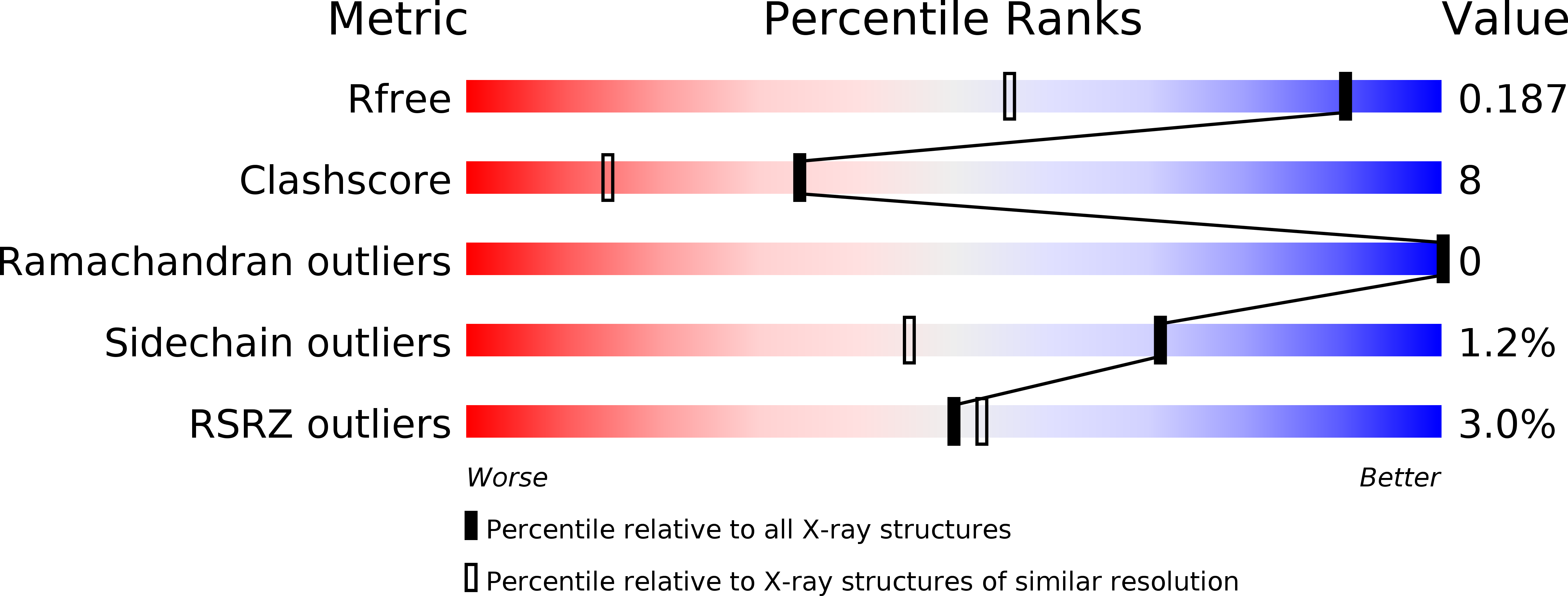
Resolution:
1.38 Å
R-Value Free:
0.17
R-Value Work:
0.15
R-Value Observed:
0.15
Space Group:
P 21 21 21
Macromolecular Entities
Structures with similar UniProt ID
Protein Blast
Polymer Type:polypeptide(L)
Molecule:GDSL-family esterase
Chain IDs:A
Chain Length:360
Number of Molecules:1
Biological Source:Photobacterium sp. J15(2011)
Primary Citation
Crystallization and structure elucidation of GDSL esterase of Photobacterium sp. J15.
Int. J. Biol. Macromol. 119 1188 1194 (2018)
PMID: 30102982 DOI: 10.1016/j.ijbiomac.2018.08.022

Abstact

GDSL esterase J15 (EstJ15) is a member of Family II of lipolytic enzyme. The enzyme was further classified in subgroup SGNH hydrolase due to the presence of highly conserve motif, Ser-Gly-Asn-His in four conserved blocks I, II, III, and V, respectively. X-ray quality crystal of EstJ15 was obtained from optimized formulation containing 0.10 M ammonium sulphate, 0.15 M sodium cacodylate trihydrate pH 6.5, and 20% PEG 8000. The crystal structure of EstJ15 was solved at 1.38 Å with one molecule per asymmetric unit. The structure exhibits α/β hydrolase fold and shared low amino acid sequence identity of 23% with the passenger domain of the autotransporter EstA of Pseudomonas aeruginosa. The active site is located at the centre of the structure, formed a narrow tunnel that hinder long substrates to be catalysed which was proven by the protein-ligand docking analysis. This study facilitates the understanding of high substrate specificity of EstJ15 and provide insights on its catalytic mechanism.

Legend

Protein

Chemical

Disease

Primary Citation of related structures
Feedback Form
Name
Email
Institute
Feedback