5XSG image
Deposition Date 2017-06-14
Release Date 2018-04-04
Last Version Date 2024-03-27
Entry Detail
PDB ID:
5XSG
Title:
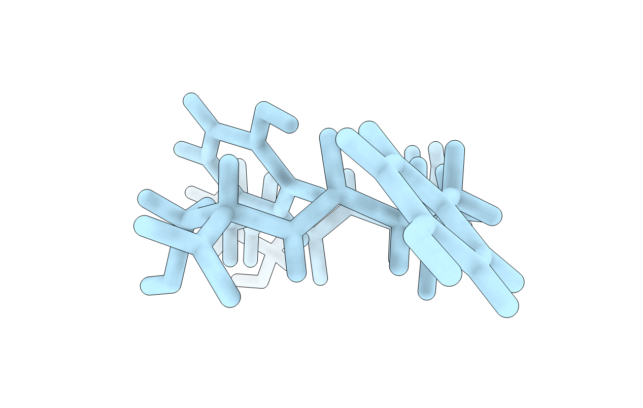
Ultrahigh resolution structure of FUS (37-42) SYSGYS determined by MicroED
Biological Source:
Source Organism:
Homo sapiens (Taxon ID: 9606)
Method Details:
Experimental Method:
Resolution:
0.73 Å
R-Value Free:
0.28
R-Value Work:
0.26
R-Value Observed:
0.26
Space Group:
P 1 21 1
Macromolecular Entities
Structures with similar UniProt ID
Protein Blast
Polymer Type:polypeptide(L)
Molecule:RNA-binding protein FUS
Gene (Uniprot):FUS
Chain IDs:A
Chain Length:6
Number of Molecules:1
Biological Source:Homo sapiens
Primary Citation
Atomic structures of FUS LC domain segments reveal bases for reversible amyloid fibril formation.
Nat. Struct. Mol. Biol. 25 341 346 (2018)
PMID: 29610493 DOI: 10.1038/s41594-018-0050-8

Abstact

Thermostable cross-β structures are characteristic of pathological amyloid fibrils, but these structures cannot explain the reversible nature of fibrils formed by RNA-binding proteins such as fused in sarcoma (FUS), involved in RNA granule assembly. Here, we find that two tandem (S/G)Y(S/G) motifs of the human FUS low-complexity domain (FUS LC) form reversible fibrils in a temperature- and phosphorylation-dependent manner. We named these motifs reversible amyloid cores, or RAC1 and RAC2, and determined their atomic structures in fibrillar forms, using microelectron and X-ray diffraction techniques. The RAC1 structure features an ordered-coil fibril spine rather than the extended β-strand typical of amyloids. Ser42, a phosphorylation site of FUS, is critical in the maintenance of the ordered-coil structure, which explains how phosphorylation controls fibril formation. The RAC2 structure shows a labile fibril spine with a wet interface. These structures illuminate the mechanism of reversible fibril formation and dynamic assembly of RNA granules.

Legend

Protein

Chemical

Disease

Primary Citation of related structures
Feedback Form
Name
Email
Institute
Feedback