
Deposition Date
2017-06-11
Release Date
2018-04-25
Last Version Date
2023-11-22
Entry Detail
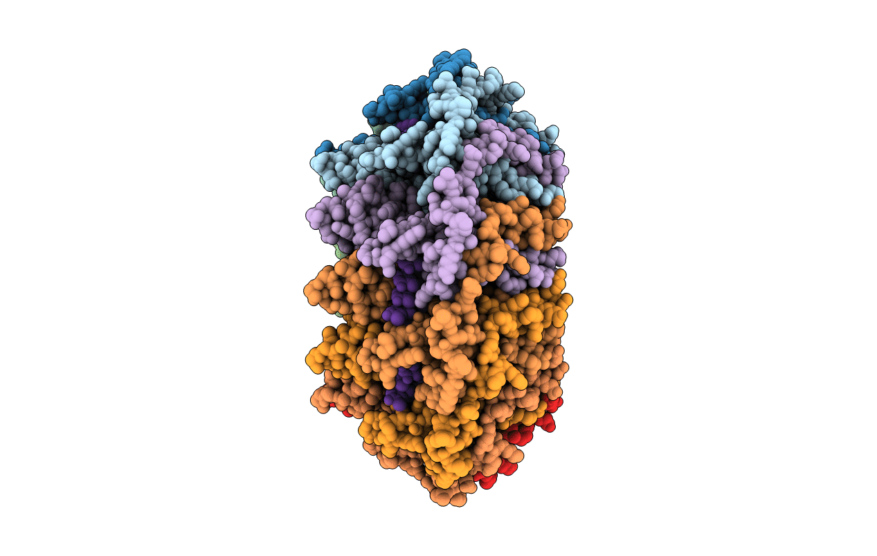
PDB ID:
5XRZ
Keywords:
Title:
Structure of a ssDNA bound to the inner DNA binding site of RAD52
Biological Source:
Source Organism(s):
Homo sapiens (Taxon ID: 9606)
synthetic construct (Taxon ID: 32630)
synthetic construct (Taxon ID: 32630)
Expression System(s):
Method Details:
Experimental Method:
Resolution:
3.60 Å
R-Value Free:
0.25
R-Value Work:
0.21
R-Value Observed:
0.21
Space Group:
P 1
