5XRW image
Deposition Date 2017-06-10
Release Date 2018-06-13
Last Version Date 2024-03-27
Entry Detail
PDB ID:
5XRW
Keywords:
Title:
Crystal structure of flagellar motor switch complex from H. pylori
Biological Source:
Source Organism:
Host Organism:
Method Details:
Experimental Method:
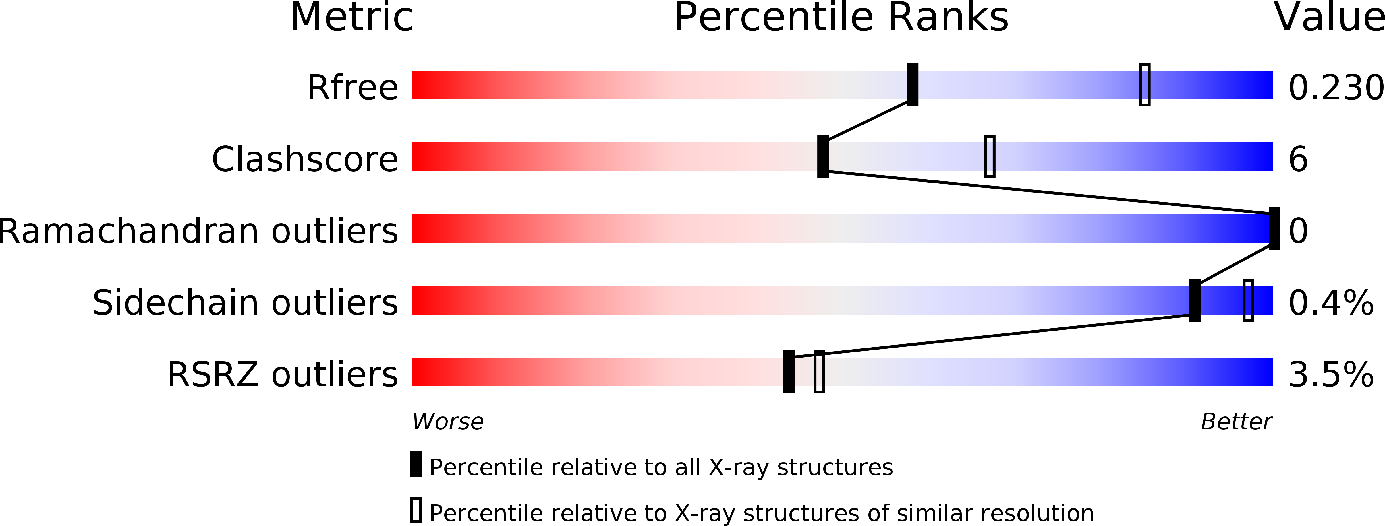
Resolution:
2.50 Å
R-Value Free:
0.22
R-Value Work:
0.20
R-Value Observed:
0.20
Space Group:
P 1
Macromolecular Entities
Structures with similar UniProt ID
Protein Blast
Polymer Type:polypeptide(L)
Molecule:FliN
Chain IDs:A, B
Chain Length:79
Number of Molecules:2
Biological Source:Helicobacter pylori 26695
Structures with similar UniProt ID
Protein Blast
Polymer Type:polypeptide(L)
Molecule:FliY
Chain IDs:C, D
Chain Length:79
Number of Molecules:2
Biological Source:Helicobacter pylori 26695
Primary Citation
Three SpoA-domain proteins interact in the creation of the flagellar type III secretion system inHelicobacter pylori.
J.Biol.Chem. 293 13961 13973 (2018)
PMID: 29991595 DOI: 10.1074/jbc.RA118.002263

Abstact

Bacterial flagella are rotary nanomachines that contribute to bacterial fitness in many settings, including host colonization. The flagellar motor relies on the multiprotein flagellar motor-switch complex to govern flagellum formation and rotational direction. Different bacteria exhibit great diversity in their flagellar motors. One such variation is exemplified by the motor-switch apparatus of the gastric pathogen Helicobacter pylori, which carries an extra switch protein, FliY, along with the more typical FliG, FliM, and FliN proteins. All switch proteins are needed for normal flagellation and motility in H. pylori, but the molecular mechanism of their assembly is unknown. To fill this gap, we examined the interactions among these proteins. We found that the C-terminal SpoA domain of FliY (FliYC) is critical to flagellation and forms heterodimeric complexes with the FliN and FliM SpoA domains, which are β-sheet domains of type III secretion system proteins. Surprisingly, unlike in other flagellar switch systems, neither FliY nor FliN self-associated. The crystal structure of the FliYC-FliNC complex revealed a saddle-shaped structure homologous to the FliN-FliN dimer of Thermotoga maritima, consistent with a FliY-FliN heterodimer forming the functional unit. Analysis of the FliYC-FliNC interface indicated that oppositely charged residues specific to each protein drive heterodimer formation. Moreover, both FliYC-FliMC and FliYC-FliNC associated with the flagellar regulatory protein FliH, explaining their important roles in flagellation. We conclude that H. pylori uses a FliY-FliN heterodimer instead of a homodimer and creates a switch complex with SpoA domains derived from three distinct proteins.

Legend

Protein

Chemical

Disease

Primary Citation of related structures
Feedback Form
Name
Email
Institute
Feedback