5XQ2 image
Deposition Date 2017-06-06
Release Date 2017-10-04
Last Version Date 2023-11-22
Entry Detail
PDB ID:
5XQ2
Title:
Crystal structure of T. thermophilus Argonaute protein complexed with a bulge 5A6 on the guide strand
Biological Source:
Method Details:
Experimental Method:
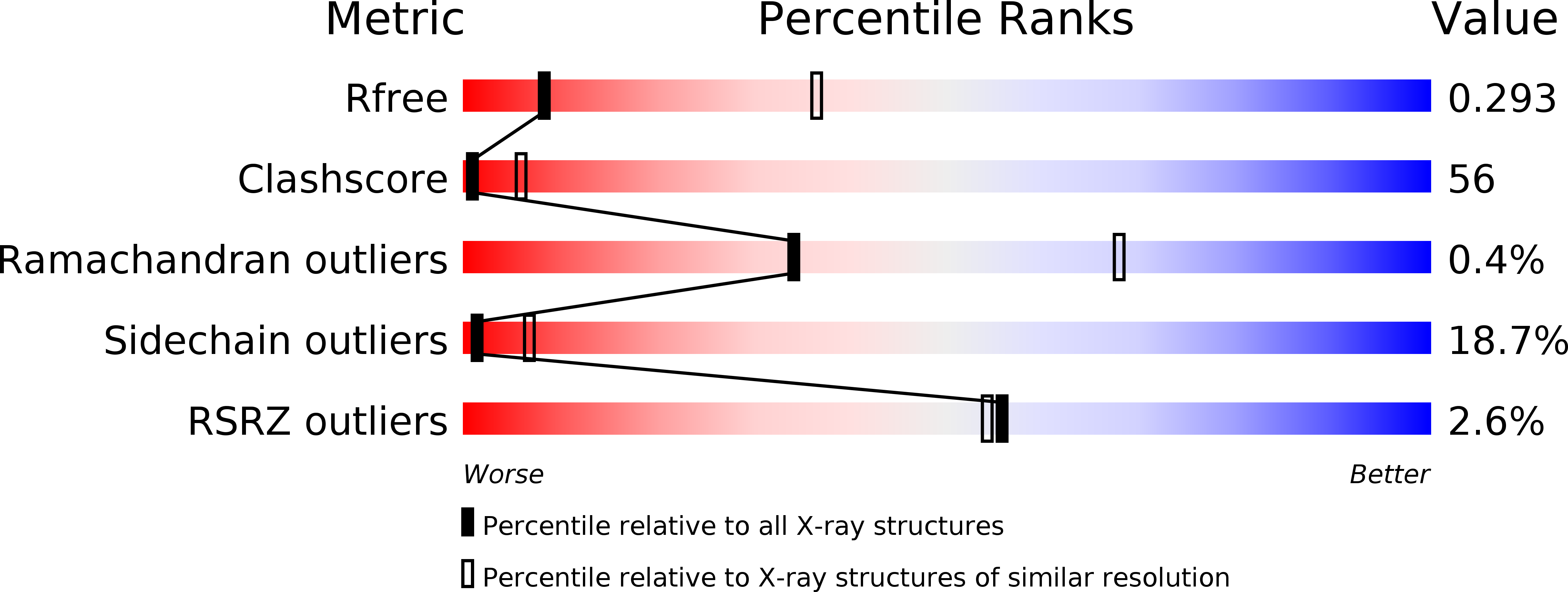
Resolution:
3.33 Å
R-Value Free:
0.29
R-Value Work:
0.25
R-Value Observed:
0.25
Space Group:
P 21 3
Macromolecular Entities
Polymer Type:polypeptide(L)
Molecule:TtAgo (D546N)
Gene (Uniprot):ago
Mutations:D546N
Chain IDs:A, E (auth: B)
Chain Length:685
Number of Molecules:2
Biological Source:Thermus thermophilus (strain HB27 / ATCC BAA-163 / DSM 7039)
Polymer Type:polydeoxyribonucleotide
Molecule:DNA (5'-D(*AP*GP*T)-3')
Chain IDs:D (auth: C), H (auth: D)
Chain Length:3
Number of Molecules:2
Biological Source:Thermus thermophilus
Polymer Type:polydeoxyribonucleotide
Molecule:DNA (5'-D(P*TP*GP*AP*AP*GP*AP*TP*AP*GP*TP*AP*GP*GP*TP*TP*GP*T)-3')
Chain IDs:B (auth: X), F (auth: E)
Chain Length:22
Number of Molecules:2
Biological Source:Thermus thermophilus
Polymer Type:polydeoxyribonucleotide
Molecule:DNA (5'-D(*AP*CP*AP*AP*CP*CP*TP*AP*CP*TP*AP*CP*CP*TP*CP*G)-3')
Chain IDs:C (auth: Y), G (auth: F)
Chain Length:19
Number of Molecules:2
Biological Source:Thermus thermophilus
Ligand Molecules
Primary Citation
Structure/cleavage-based insights into helical perturbations at bulge sites within T. thermophilus Argonaute silencing complexes
Nucleic Acids Res. 45 9149 9163 (2017)
PMID: 28911094 DOI: 10.1093/nar/gkx547

Abstact

We have undertaken a systematic structural study of Thermus thermophilus Argonaute (TtAgo) ternary complexes containing single-base bulges positioned either within the seed segment of the guide or target strands and at the cleavage site. Our studies establish that single-base bulges 7T8, 5A6 and 4A5 on the guide strand are stacked-into the duplex, with conformational changes localized to the bulge site, thereby having minimal impact on the cleavage site. By contrast, single-base bulges 6'U7' and 6'A7' on the target strand are looped-out of the duplex, with the resulting conformational transitions shifting the cleavable phosphate by one step. We observe a stable alignment for the looped-out 6'N7' bulge base, which stacks on the unpaired first base of the guide strand, with the looped-out alignment facilitated by weakened Watson-Crick and reversed non-canonical flanking pairs. These structural studies are complemented by cleavage assays that independently monitor the impact of bulges on TtAgo-mediated cleavage reaction.

Legend

Protein

Chemical

Disease

Primary Citation of related structures