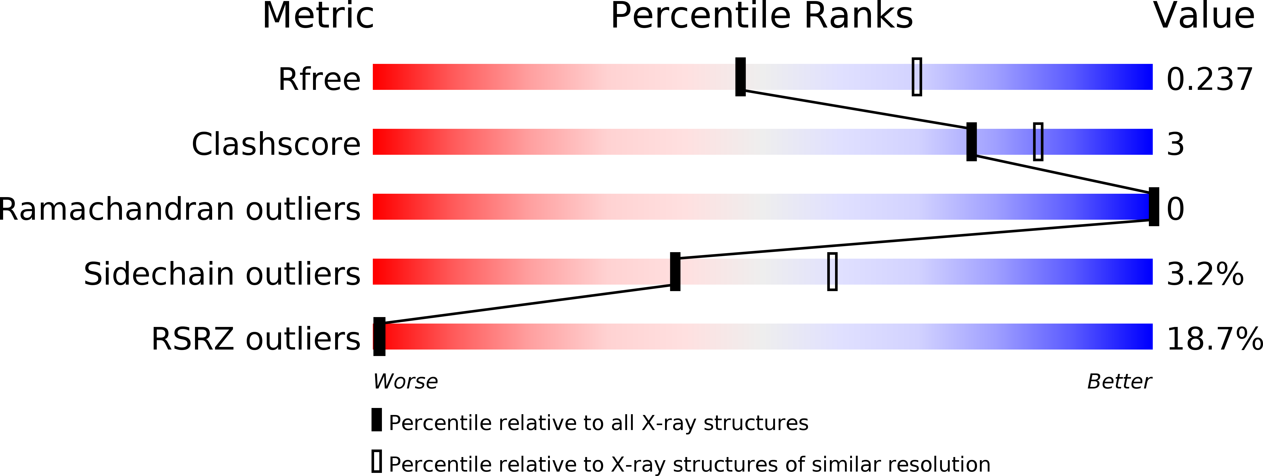
5XOC image
Deposition Date 2017-05-27
Release Date 2018-03-28
Last Version Date 2024-10-16
Entry Detail
PDB ID:
5XOC
Keywords:
Title:
Crystal structure of human Smad3-FoxH1 complex
Biological Source:
Source Organism:
Host Organism:
Method Details:
Experimental Method:
Resolution:
2.40 Å
R-Value Free:
0.23
R-Value Work:
0.19
R-Value Observed:
0.19
Space Group:
P 63 2 2
Macromolecular Entities
Polymer Type:polypeptide(L)
Molecule:Mothers against decapentaplegic homolog 3
Gene (Uniprot):SMAD3
Chain IDs:A
Chain Length:199
Number of Molecules:1
Biological Source:Homo sapiens
Polymer Type:polypeptide(L)
Molecule:Thioredoxin 1,Forkhead box protein H1
Gene (Uniprot):FOXH1, trxA
Chain IDs:B
Chain Length:141
Number of Molecules:1
Biological Source:Escherichia coli (strain K12), Homo sapiens
Primary Citation
Hydrophobic patches on SMAD2 and SMAD3 determine selective binding to cofactors
Sci Signal 11 ? ? (2018)
PMID: 29588413 DOI: 10.1126/scisignal.aao7227

Abstact

The transforming growth factor-β (TGF-β) superfamily of cytokines regulates various biological processes, including cell proliferation, immune responses, autophagy, and senescence. Dysregulation of TGF-β signaling causes various diseases, such as cancer and fibrosis. SMAD2 and SMAD3 are core transcription factors involved in TGF-β signaling, and they form heterotrimeric complexes with SMAD4 (SMAD2-SMAD2-SMAD4, SMAD3-SMAD3-SMAD4, and SMAD2-SMAD3-SMAD4) in response to TGF-β signaling. These heterotrimeric complexes interact with cofactors to control the expression of TGF-β-dependent genes. SMAD2 and SMAD3 may promote or repress target genes depending on whether they form complexes with other transcription factors, coactivators, or corepressors; therefore, the selection of specific cofactors is critical for the appropriate activity of these transcription factors. To reveal the structural basis by which SMAD2 and SMAD3 select cofactors, we determined the crystal structures of SMAD3 in complex with the transcription factor FOXH1 and SMAD2 in complex with the transcriptional corepressor SKI. The structures of the complexes show that the MAD homology 2 (MH2) domains of SMAD2 and SMAD3 have multiple hydrophobic patches on their surfaces. The cofactors tether to various subsets of these patches to interact with SMAD2 and SMAD3 in a cooperative or competitive manner to control the output of TGF-β signaling.

Legend

Protein

Chemical

Disease

Primary Citation of related structures