5XNX image
Deposition Date 2017-05-24
Release Date 2017-07-19
Last Version Date 2023-11-22
Entry Detail
PDB ID:
5XNX
Title:
Crystallographic structure of the enzymatically active N-terminal domain of the Rel protein from Mycobacterium tuberculosis
Biological Source:
Method Details:
Experimental Method:
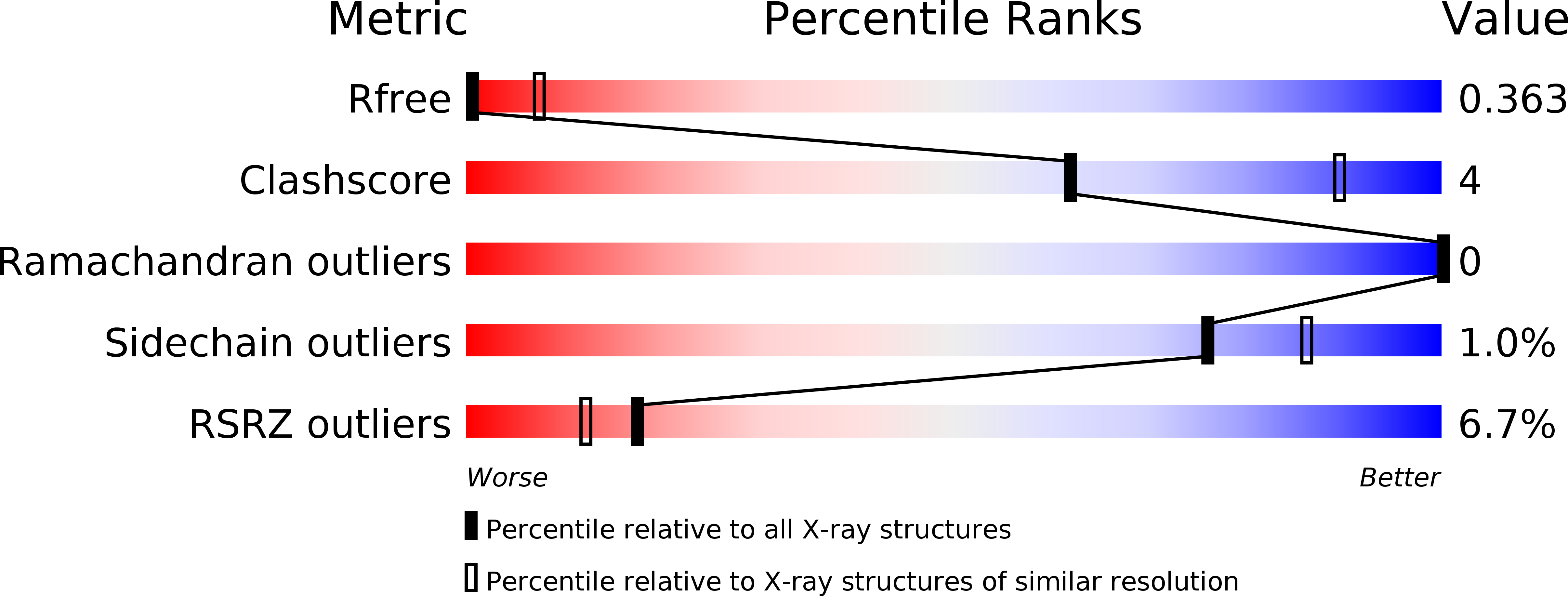
Resolution:
3.70 Å
R-Value Free:
0.36
R-Value Work:
0.35
R-Value Observed:
0.35
Space Group:
P 32
Macromolecular Entities
Polymer Type:polypeptide(L)
Molecule:Bifunctional (p)ppGpp synthase/hydrolase RelA
Gene (Uniprot):relA
Chain IDs:A, B, C, D
Chain Length:394
Number of Molecules:4
Biological Source:Mycobacterium tuberculosis (strain ATCC 25618 / H37Rv)
Ligand Molecules
Primary Citation
Crystallographic and solution structure of the N-terminal domain of the Rel protein from Mycobacterium tuberculosis
FEBS Lett. 591 2323 2337 (2017)
PMID: 28672070 DOI: 10.1002/1873-3468.12739

Abstact

Modulation of intracellular guanosine 3',5'-bispyrophosphate ((p)ppGpp) level, the effector of the stringent response, is crucial for survival as well as optimal growth of prokaryotes and, thus, for bacterial pathogenesis and dormancy. In Mycobacterium tuberculosis (Mtb), (p)ppGpp synthesis and degradation are carried out by the bifunctional enzyme MtRel, which consists of 738 residues, including an N-terminal hydrolase- and synthetase-domain (N-terminal domain or NTD) and a C-terminus with a ribosome-binding site. Here, we present the first crystallographic structure of the enzymatically active MtRel NTD determined at 3.7 Å resolution. The structure provides insights into the residues of MtRel NTD responsible for nucleotide binding. Small-angle X-ray scattering experiments were performed to investigate the dimeric state of the MtRel NTD and possible substrate-dependent structural alterations.

Legend

Protein

Chemical

Disease

Primary Citation of related structures