5XGR image
Deposition Date 2017-04-16
Release Date 2017-05-10
Last Version Date 2024-10-09
Entry Detail
PDB ID:
5XGR
Keywords:
Title:
Structure of the S1 subunit C-terminal domain from bat-derived coronavirus HKU5 spike protein
Biological Source:
Source Organism:
Host Organism:
Method Details:
Experimental Method:
Resolution:
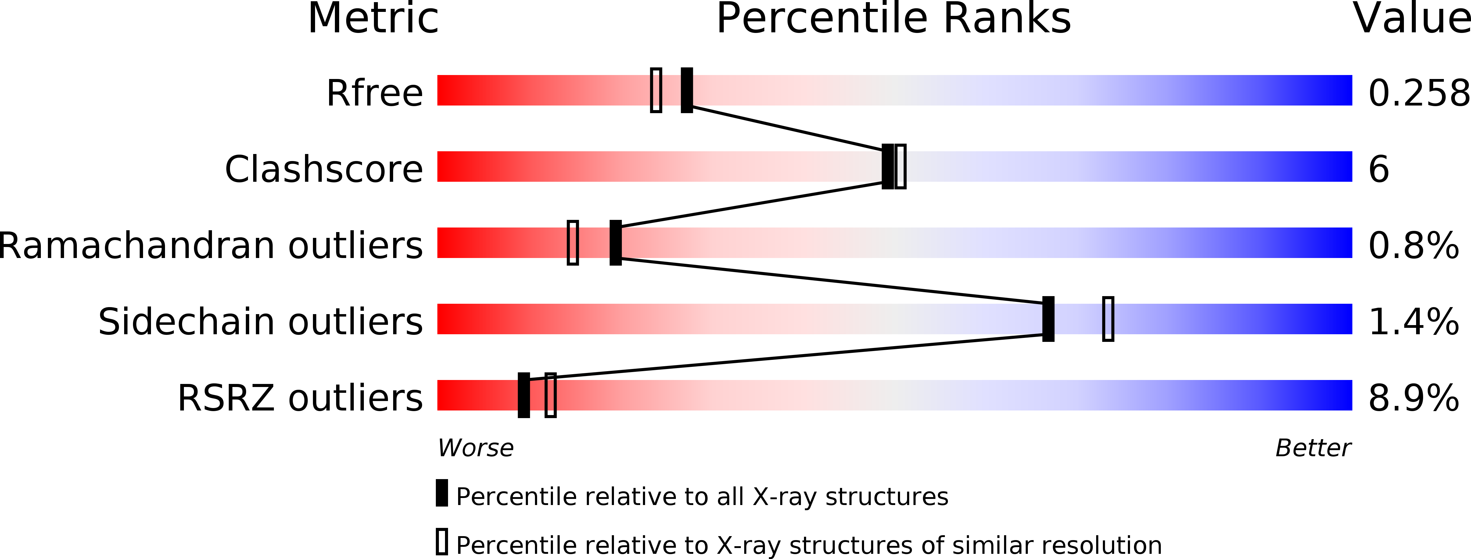
2.10 Å
R-Value Free:
0.25
R-Value Work:
0.21
R-Value Observed:
0.21
Space Group:
P 1 21 1
Macromolecular Entities
Structures with similar UniProt ID
Protein Blast
Polymer Type:polypeptide(L)
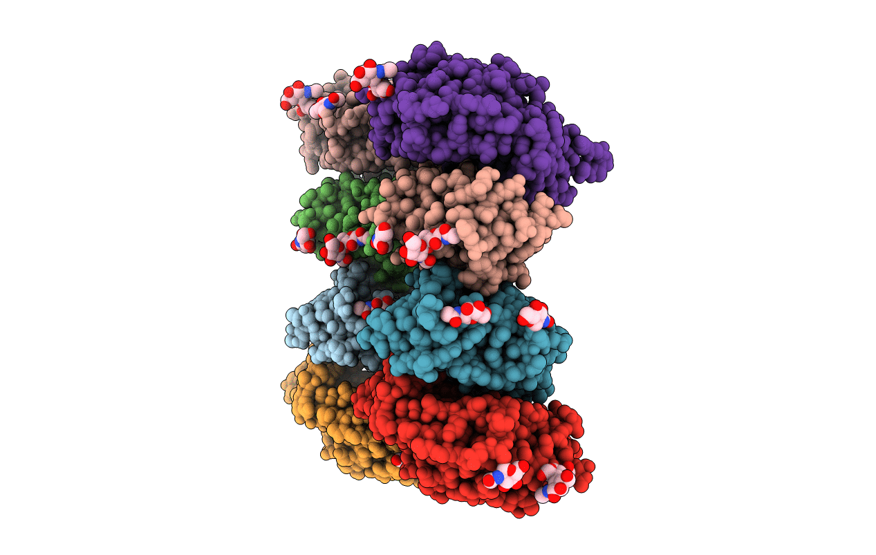
Molecule:Spike protein S1
Gene (Uniprot):S
Chain IDs:A, B, C, D, E, F, G, H
Chain Length:204
Number of Molecules:8
Biological Source:Bat coronavirus HKU5
Ligand Molecules
Primary Citation
Structure of the S1 subunit C-terminal domain from bat-derived coronavirus HKU5 spike protein
Virology 507 101 109 (2017)
PMID: 28432925 DOI: 10.1016/j.virol.2017.04.016

Abstact

Accumulating evidence indicates that MERS-CoV originated from bat coronaviruses (BatCoVs). Previously, we demonstrated that both MERS-CoV and BatCoV HKU4 use CD26 as a receptor, but how the BatCoVs evolved to bind CD26 is an intriguing question. Here, we solved the crystal structure of the S1 subunit C-terminal domain of HKU5 (HKU5-CTD), another BatCoV that is phylogenetically related to MERS-CoV but cannot bind to CD26. We observed that the conserved core subdomain and those of other betacoronaviruses (betaCoVs) have a similar topology of the external subdomain, indicating the same ancestor of lineage C betaCoVs. However, two deletions in two respective loops located in HKU5-CTD result in conformational variations in CD26-binding interface and are responsible for the non-binding of HKU5-CTD to CD26. Combined with sequence variation in the HKU5-CTD receptor binding interface, we propose the necessity for surveilling the mutation in BatCoV HKU5 spike protein in case of bat-to-human interspecies transmission.

Legend

Protein

Chemical

Disease

Primary Citation of related structures