5XFW image
Deposition Date 2017-04-11
Release Date 2018-04-25
Last Version Date 2023-11-22
Entry Detail
PDB ID:
5XFW
Keywords:
Title:
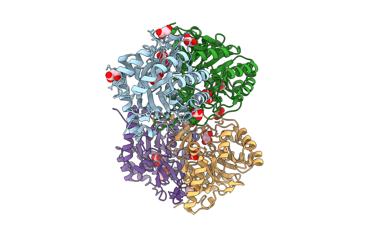
Crystal structures of FMN-free form of dihydroorotate dehydrogenase from Trypanosoma brucei
Biological Source:
Host Organism:
Method Details:
Experimental Method:
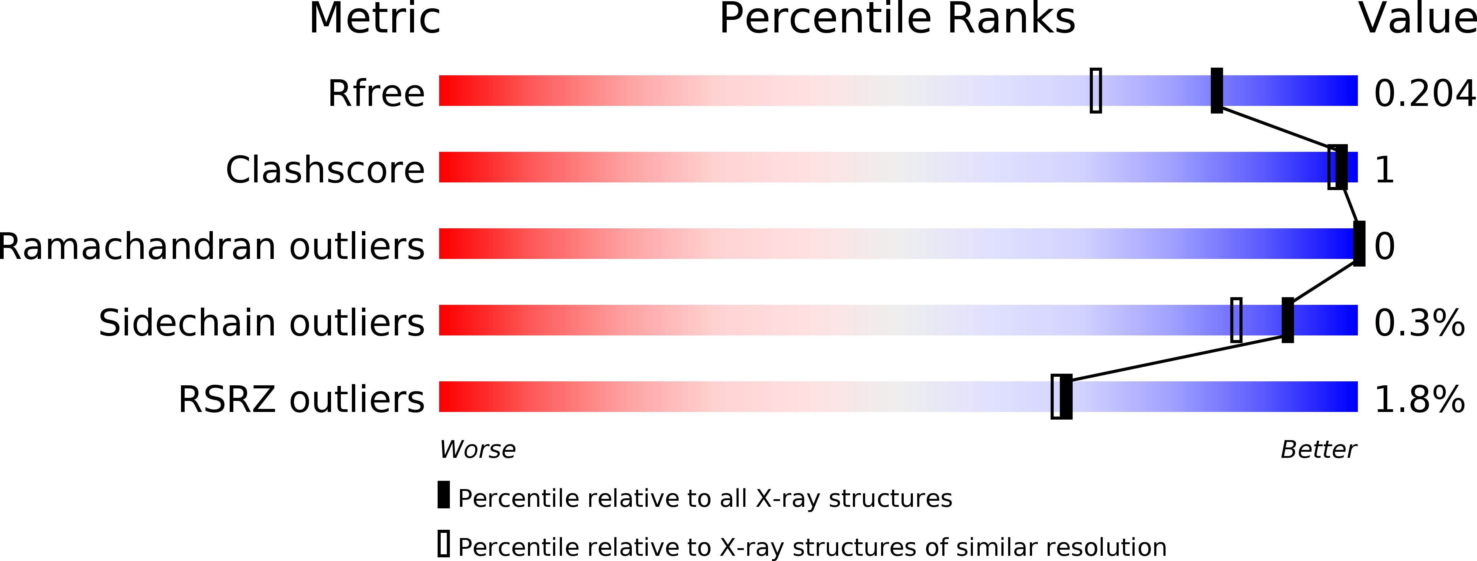
Resolution:
1.60 Å
R-Value Free:
0.19
R-Value Work:
0.17
R-Value Observed:
0.17
Space Group:
P 21 21 2
Macromolecular Entities
Polymer Type:polypeptide(L)
Molecule:Dihydroorotate dehydrogenase (fumarate)
Gene (Uniprot):Tb927.5.3830
Mutations:A115V
Chain IDs:A, B, C, D
Chain Length:334
Number of Molecules:4
Biological Source:Trypanosoma brucei brucei (strain 927/4 GUTat10.1)
Ligand Molecules
Primary Citation
Crystal structures of FMN-bound and FMN-free forms of dihydroorotate dehydrogenase fromTrypanosoma brucei.
FEBS Open Bio 8 680 691 (2018)
PMID: 29632820 DOI: 10.1002/2211-5463.12403

Abstact

Dihydroorotate dehydrogenase (DHODH) is a flavin-binding enzyme essential for pyrimidine biosynthesis, which converts dihydroorotate to orotate. Three-dimensional structures of cytosolic DHODH of parasitic protozoa are of interest in drug discovery for neglected tropical diseases, especially because these enzymes possess significantly different structural and functional properties from the membrane-associated human enzyme. The existing crystal structures of the flavin mononucleotide (FMN)-bound DHODHs reveal a number of interactions stabilizing FMN. However, to understand the binding mechanism correctly, it is necessary to compare the structures of the FMN-bound and FMN-free forms, because the protein moiety of the former is not necessarily the same as the latter. Here, we prepared the FMN-free DHODH of Trypanosoma brucei using an Escherichia coli overexpression system. Although this apoform lacks enzymatic activity, simple incubation with FMN activated the enzyme. It was stable enough to be crystallized, enabling us to determine its structure by X-ray crystallography at 1.6 Å resolution. We also determined the FMN-bound form at 1.8 Å resolution. Although the two structures have essentially the same scaffold, we observed flipping of a peptide-bond plane in the vicinity of the FMN-binding site, accompanied by an alternative hydrogen-bonding pattern. Comparisons of B factors of the protein main chain revealed that binding of FMN decreased flexibility of most of the residues at the FMN-binding site, but increased flexibility of a lid-like loop structure over the active center. This increase was ascribed to a conformational change in an FMN-contacting residue, Asn195, which induced a rearrangement of a hydrogen-bond network of the residues comprising the lid.

Legend

Protein

Chemical

Disease

Primary Citation of related structures