5XFS image
Deposition Date 2017-04-11
Release Date 2017-08-30
Last Version Date 2023-11-22
Entry Detail
PDB ID:
5XFS
Title:
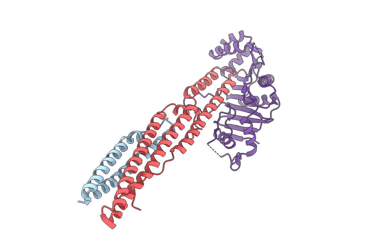
Crystal structure of PE8-PPE15 in complex with EspG5 from M. tuberculosis
Biological Source:
Method Details:
Experimental Method:
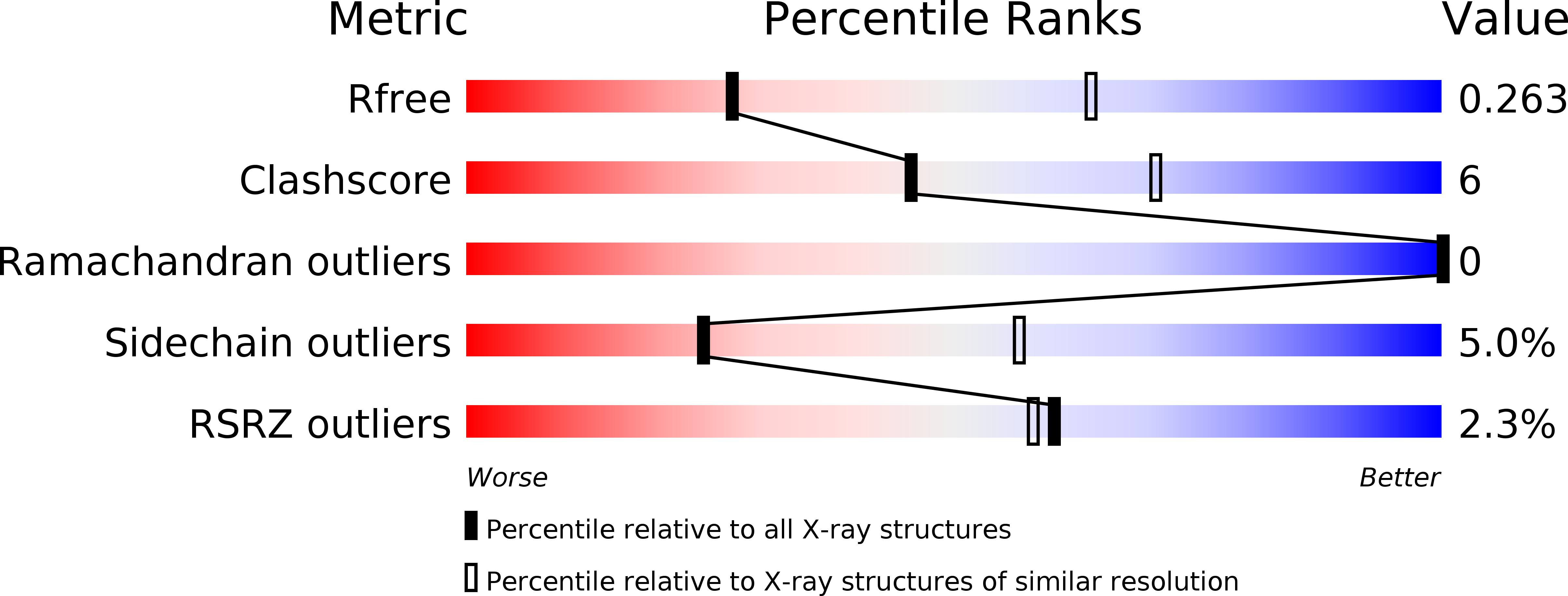
Resolution:
2.90 Å
R-Value Free:
0.26
R-Value Work:
0.21
R-Value Observed:
0.21
Space Group:
P 21 21 21
Macromolecular Entities
Structures with similar UniProt ID
Protein Blast
Polymer Type:polypeptide(L)
Molecule:PE family protein PE8
Gene (Uniprot):PE8
Chain IDs:A
Chain Length:104
Number of Molecules:1
Biological Source:Mycobacterium tuberculosis (strain ATCC 25618 / H37Rv)
Structures with similar UniProt ID
Protein Blast
Polymer Type:polypeptide(L)
Molecule:PPE family protein PPE15
Gene (Uniprot):PPE15
Chain IDs:B
Chain Length:202
Number of Molecules:1
Biological Source:Mycobacterium tuberculosis (strain ATCC 25618 / H37Rv)
Structures with similar UniProt ID
Protein Blast
Polymer Type:polypeptide(L)
Molecule:ESX-5 secretion-associated protein EspG5
Gene (Uniprot):espG5
Chain IDs:C
Chain Length:308
Number of Molecules:1
Biological Source:Mycobacterium tuberculosis (strain ATCC 25618 / H37Rv)
Primary Citation
Structural basis of the PE-PPE protein interaction in Mycobacterium tuberculosis.
J. Biol. Chem. 292 16880 16890 (2017)
PMID: 28842489 DOI: 10.1074/jbc.M117.802645

Abstact

Mycobacterium tuberculosis (Mtb), the causative agent of tuberculosis, has developed multiple strategies to adapt to the human host. The five type VII secretion systems, ESX-1-5, direct the export of many virulence-promoting protein effectors across the complex mycobacterial cell wall. One class of ESX substrates is the PE-PPE family of proteins, which is unique to mycobacteria and essential for infection, antigenic variation, and host-pathogen interactions. The genome of Mtb encodes 168 PE-PPE proteins. Many of them are thought to be secreted through ESX-5 secretion system and to function in pairs. However, understanding of the specific pairing of PE-PPE proteins and their structure-function relationship is limited by the challenging purification of many PE-PPE proteins, and our knowledge of the PE-PPE interactions therefore has been restricted to the PE25-PPE41 pair and its complex with the ESX-5 secretion system chaperone EspG5. Here, we report the crystal structure of a new PE-PPE pair, PE8-PPE15, in complex with EspG5. Our structure revealed that the EspG5-binding sites on PPE15 are relatively conserved among Mtb PPE proteins, suggesting that EspG5-PPE15 represents a more typical model for EspG5-PPE interactions than EspG5-PPE41. A structural comparison with the PE25-PPE41 complex disclosed conformational changes in the four-helix bundle structure and a unique binding mode in the PE8-PPE15 pair. Moreover, homology-modeling and mutagenesis studies further delineated the molecular determinants of the specific PE-PPE interactions. These findings help develop an atomic algorithm of ESX-5 substrate recognition and PE-PPE pairing.

Legend

Protein

Chemical

Disease

Primary Citation of related structures