Deposition Date
2017-04-10
Release Date
2017-10-18
Last Version Date
2023-11-22
Entry Detail
PDB ID:
5XFI
Keywords:
Title:
Crystal structure of Calsepa lectin in complex with biantennary N-glycan
Biological Source:
Source Organism(s):
Calystegia sepium (Taxon ID: 47519)
Expression System(s):
Method Details:
Experimental Method:
Resolution:
1.65 Å
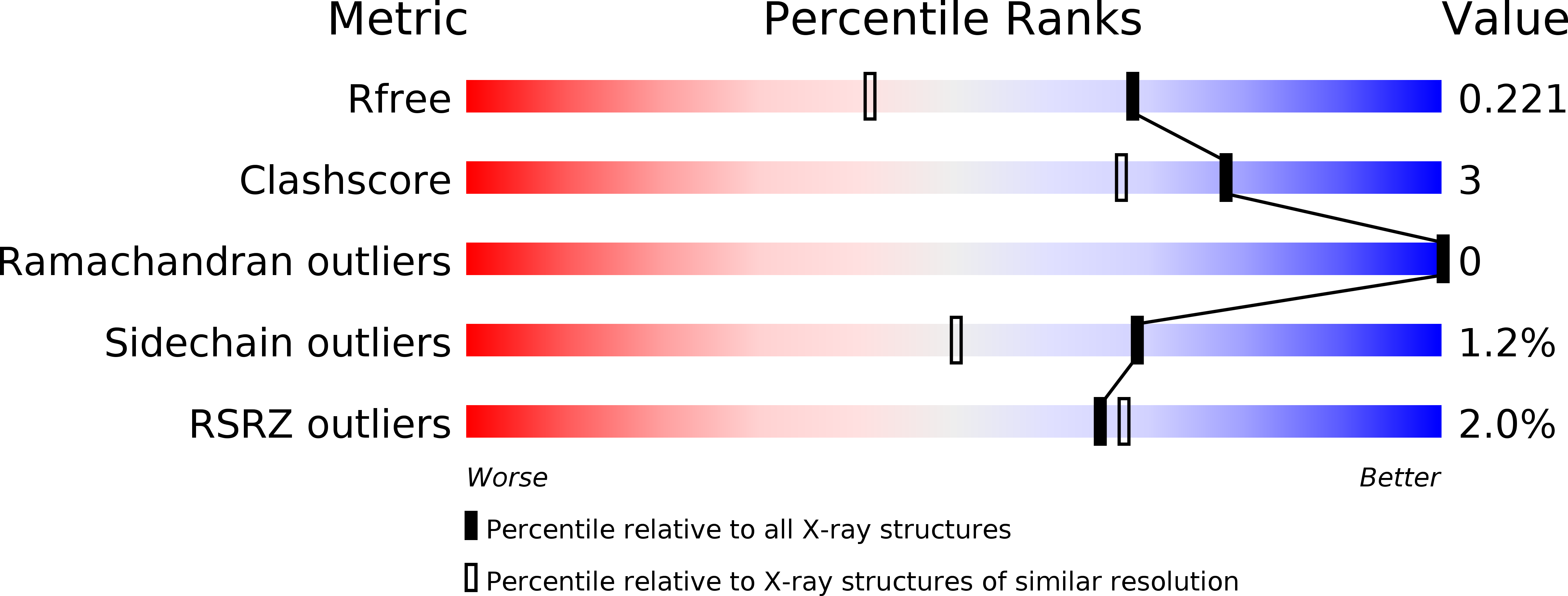
R-Value Free:
0.22
R-Value Work:
0.18
R-Value Observed:
0.18
Space Group:
P 43