Deposition Date
2017-04-08
Release Date
2017-12-13
Last Version Date
2023-11-22
Entry Detail
PDB ID:
5XF7
Keywords:
Title:
Crystal structure of human protein disulfide isomerase-like protein of the testis
Biological Source:
Source Organism(s):
Homo sapiens (Taxon ID: 9606)
Expression System(s):
Method Details:
Experimental Method:
Resolution:
2.38 Å
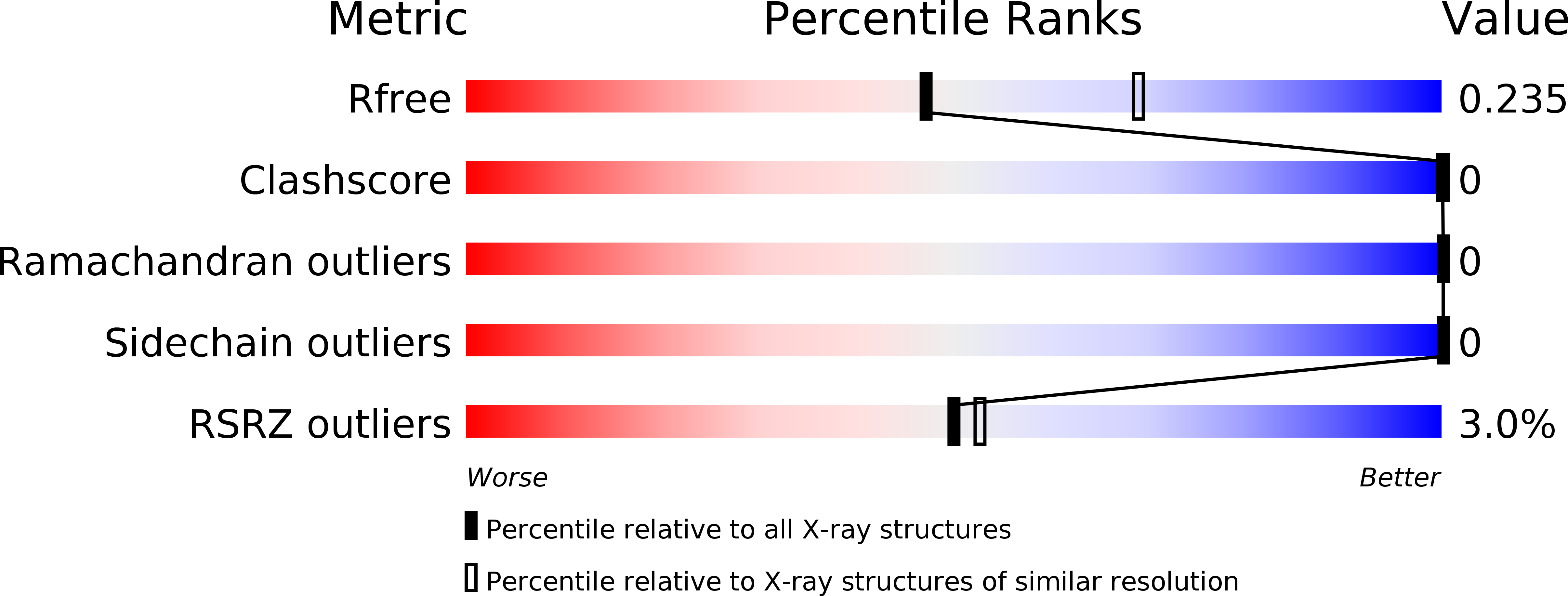
R-Value Free:
0.23
R-Value Work:
0.19
R-Value Observed:
0.20
Space Group:
P 32