5XE3 image
Deposition Date 2017-03-31
Release Date 2017-10-11
Last Version Date 2023-11-22
Entry Detail
PDB ID:
5XE3
Title:
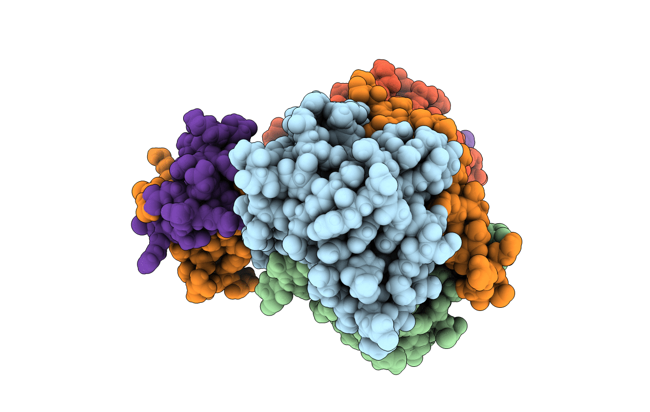
Endoribonuclease in complex with its cognate antitoxin from Mycobacterial species
Biological Source:
Method Details:
Experimental Method:
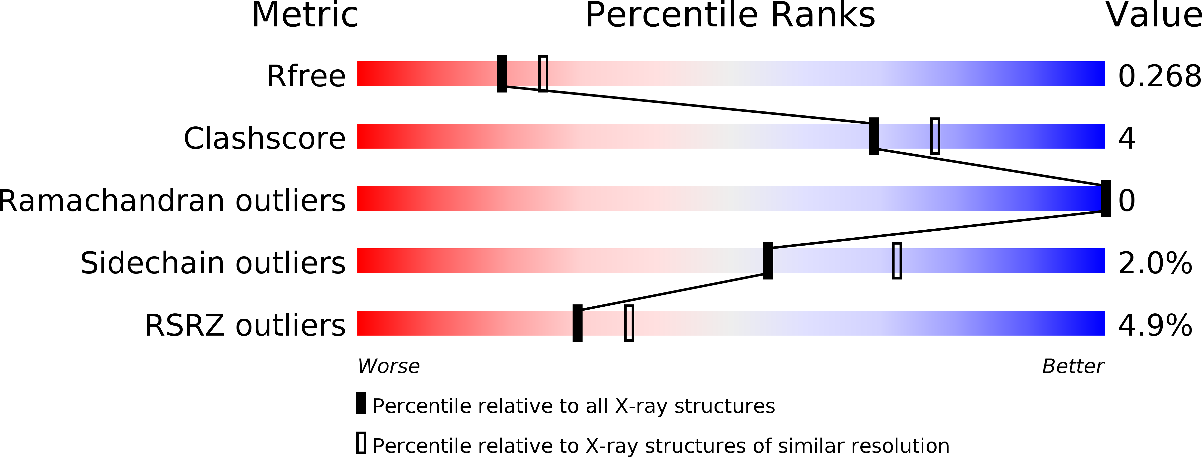
Resolution:
2.30 Å
R-Value Free:
0.26
R-Value Work:
0.21
R-Value Observed:
0.21
Space Group:
P 1 21 1
Macromolecular Entities
Structures with similar UniProt ID
Protein Blast
Polymer Type:polypeptide(L)
Molecule:Endoribonuclease MazF4
Gene (Uniprot):mazF4
Chain IDs:A (auth: C), B (auth: D), D (auth: A), E (auth: B)
Chain Length:107
Number of Molecules:4
Biological Source:Mycobacterium tuberculosis (strain ATCC 25618 / H37Rv)
Structures with similar UniProt ID
Protein Blast
Polymer Type:polypeptide(L)
Molecule:Probable antitoxin MazE4
Gene (Uniprot):mazE4
Chain IDs:C (auth: F), F (auth: E)
Chain Length:81
Number of Molecules:2
Biological Source:Mycobacterium tuberculosis (strain ATCC 25618 / H37Rv)
Primary Citation
Structural analyses of the MazEF4 toxin-antitoxin pair in Mycobacterium tuberculosis provide evidence for a unique extracellular death factor.
J. Biol. Chem. 292 18832 18847 (2017)
PMID: 28972145 DOI: 10.1074/jbc.M117.807974

Abstact

The bacterial toxin-antitoxin MazEF system in the tuberculosis (TB)-causing bacterium Mycobacterium tuberculosis is activated under unfavorable conditions, including starvation, antibiotic exposure, and oxidative stress. This system contains the ribonucleolytic enzyme MazF and has emerged as a promising drug target for TB treatments targeting the latent stage of M. tuberculosis infection and reportedly mediates a cell death process via a peptide called extracellular death factor (EDF). Although it is well established that the increase in EDF-mediated toxicity of MazF drives a cell-killing phenomenon, the molecular details are poorly understood. Moreover, the divergence in sequences among reported EDFs suggests that each bacterial species has a unique EDF. To address these open questions, we report here the structures of MazF4 and MazEF4 complexes from M. tuberculosis, representing the first MazEF structures from this organism. We found that MazF4 possesses a negatively charged MazE4-binding pocket in contrast to the positively charged MazE-binding pockets in homologous MazEF complex structures from other bacteria. Moreover, using NMR spectroscopy and biochemical assays, we unraveled the molecular interactions of MazF4 with its RNA substrate and with a new EDF homolog originating from M. tuberculosis The EDF homolog discovered here possesses a positively charged residue at the C terminus, making this EDF distinct from previously reported EDFs. Overall, our results suggest that M. tuberculosis evolved a unique MazF and EDF and that the distinctive EDF sequence could serve as a starting point for designing new anti-tuberculosis drugs. We therefore conclude that this study might contribute to the development of a new line of anti-tuberculosis agents.

Legend

Protein

Chemical

Disease

Primary Citation of related structures