5XCY image
Deposition Date 2017-03-24
Release Date 2017-07-26
Last Version Date 2024-11-13
Entry Detail
PDB ID:
5XCY
Keywords:
Title:
Structure of the cellobiohydrolase Cel6A from Phanerochaete chrysosporium at 1.2 angstrom
Biological Source:
Source Organism:
Host Organism:
Method Details:
Experimental Method:
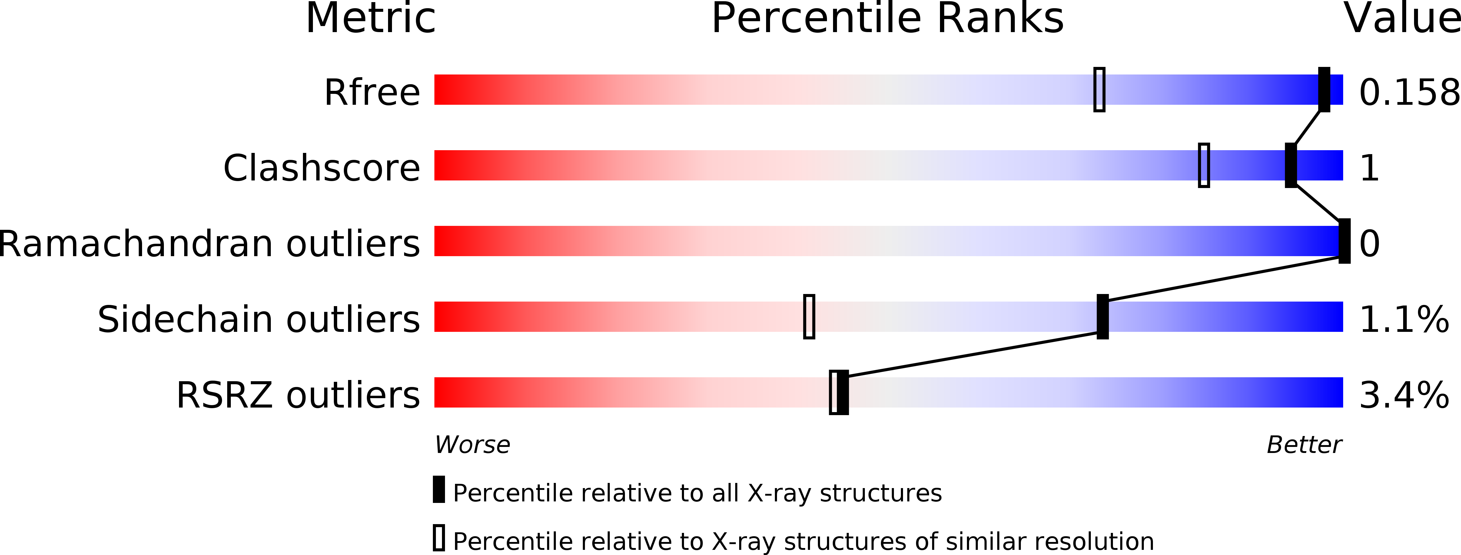
Resolution:
1.20 Å
R-Value Free:
0.15
R-Value Work:
0.13
R-Value Observed:
0.13
Space Group:
P 21 21 21
Macromolecular Entities
Structures with similar UniProt ID
Protein Blast
Polymer Type:polypeptide(L)
Molecule:Glucanase
Chain IDs:A
Chain Length:358
Number of Molecules:1
Biological Source:Phanerochaete chrysosporium
Primary Citation
Crystal structure of a family 6 cellobiohydrolase from the basidiomycete Phanerochaete chrysosporium
Acta Crystallogr F Struct Biol Commun 73 398 403 (2017)
PMID: 28695848 DOI: 10.1107/S2053230X17008093

Abstact

Cellobiohydrolases belonging to glycoside hydrolase family 6 (CBH II, Cel6A) play key roles in the hydrolysis of crystalline cellulose. CBH II from the white-rot fungus Phanerochaete chrysosporium (PcCel6A) consists of a catalytic domain (CD) and a carbohydrate-binding module connected by a linker peptide, like other known fungal cellobiohydrolases. In the present study, the CD of PcCel6A was crystallized without ligands, and p-nitrophenyl β-D-cellotrioside (pNPG3) was soaked into the crystals. The determined structures of the ligand-free and pNPG3-soaked crystals revealed that binding of cellobiose at substrate subsites +1 and +2 induces a conformational change of the N-terminal and C-terminal loops, switching the tunnel-shaped active site from the open to the closed form.

Legend

Protein

Chemical

Disease

Primary Citation of related structures