5XB7 image
Deposition Date 2017-03-16
Release Date 2017-11-01
Last Version Date 2023-11-22
Entry Detail
PDB ID:
5XB7
Keywords:
Title:
GH42 alpha-L-arabinopyranosidase from Bifidobacterium animalis subsp. lactis Bl-04
Biological Source:
Source Organism:
Host Organism:
Method Details:
Experimental Method:
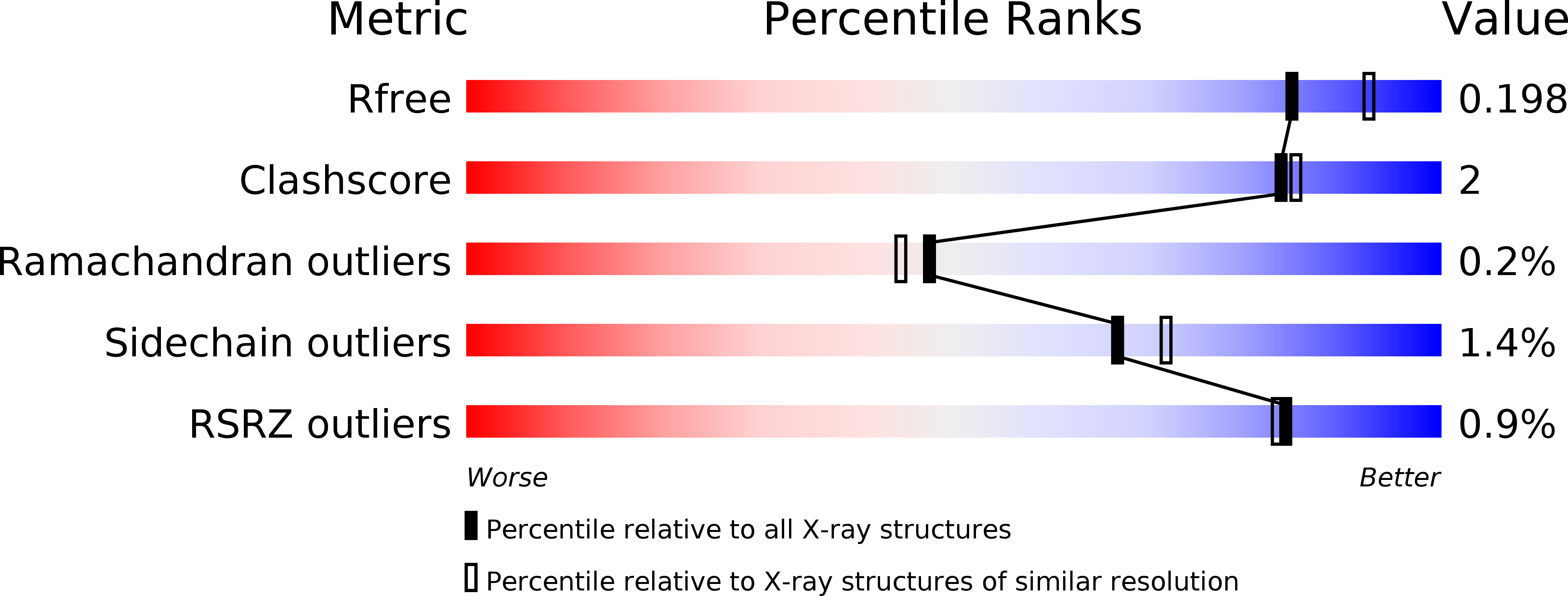
Resolution:
2.00 Å
R-Value Free:
0.19
R-Value Work:
0.15
R-Value Observed:
0.15
Space Group:
P 41 21 2
Macromolecular Entities
Polymer Type:polypeptide(L)
Molecule:Beta-galactosidase
Chain IDs:A, B, C, D, E, F
Chain Length:712
Number of Molecules:6
Biological Source:Bifidobacterium animalis subsp. lactis
Primary Citation
Discovery of alpha-l-arabinopyranosidases from human gut microbiome expands the diversity within glycoside hydrolase family 42.
J. Biol. Chem. 292 21092 21101 (2017)
PMID: 29061847 DOI: 10.1074/jbc.M117.792598

Abstact

Enzymes of the glycoside hydrolase family 42 (GH42) are widespread in bacteria of the human gut microbiome and play fundamental roles in the decomposition of both milk and plant oligosaccharides. All GH42 enzymes characterized so far have β-galactosidase activity. Here, we report the existence of a GH42 subfamily that is exclusively specific for α-l-arabinopyranoside and describe the first representative of this subfamily. We found that this enzyme (BlArap42B) from a probiotic Bifidobacterium species cannot hydrolyze β-galactosides. However, BlArap42B effectively hydrolyzed paeonolide and ginsenoside Rb2, plant glycosides containing an aromatic aglycone conjugated to α-l-arabinopyranosyl-(1,6)-β-d-glucopyranoside. Paeonolide, a natural glycoside from the roots of the plant genus Paeonia, is not hydrolyzed by classical GH42 β-galactosidases. X-ray crystallography revealed a unique Trp345-X12-Trp358 sequence motif at the BlArap42B active site, as compared with a Phe-X12-His motif in classical GH42 β-galactosidases. This analysis also indicated that the C6 position of galactose is blocked by the aromatic side chains, hence allowing accommodation only of Arap lacking this carbon. Automated docking of paeonolide revealed that it can fit into the BlArap42B active site. The Glcp moiety of paeonolide stacks onto the aromatic ring of the Trp252 at subsite +1 and C4-OH is hydrogen bonded with Asp249 Moreover, the aglycone stacks against Phe421 from the neighboring monomer in the BlArap42B trimer, forming a proposed subsite +2. These results further support the notion that evolution of metabolic specialization can be tracked at the structural level in key enzymes facilitating degradation of specific glycans in an ecological niche.

Legend

Protein

Chemical

Disease

Primary Citation of related structures