5XB2 image
Entry Detail
PDB ID:
5XB2
Keywords:
Title:
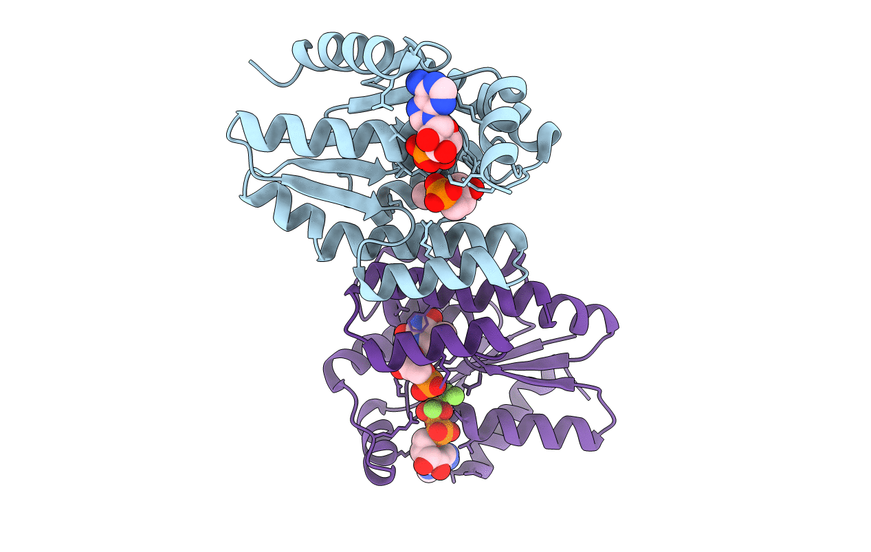
ADP-Mg-F-dTMP bound Crystal structure of thymidylate kinase (aq_969) from Aquifex Aeolicus VF5
Biological Source:
Host Organism:
PDB Version:
Deposition Date:
2017-03-15
Release Date:
2017-06-28
Method Details:
Experimental Method:
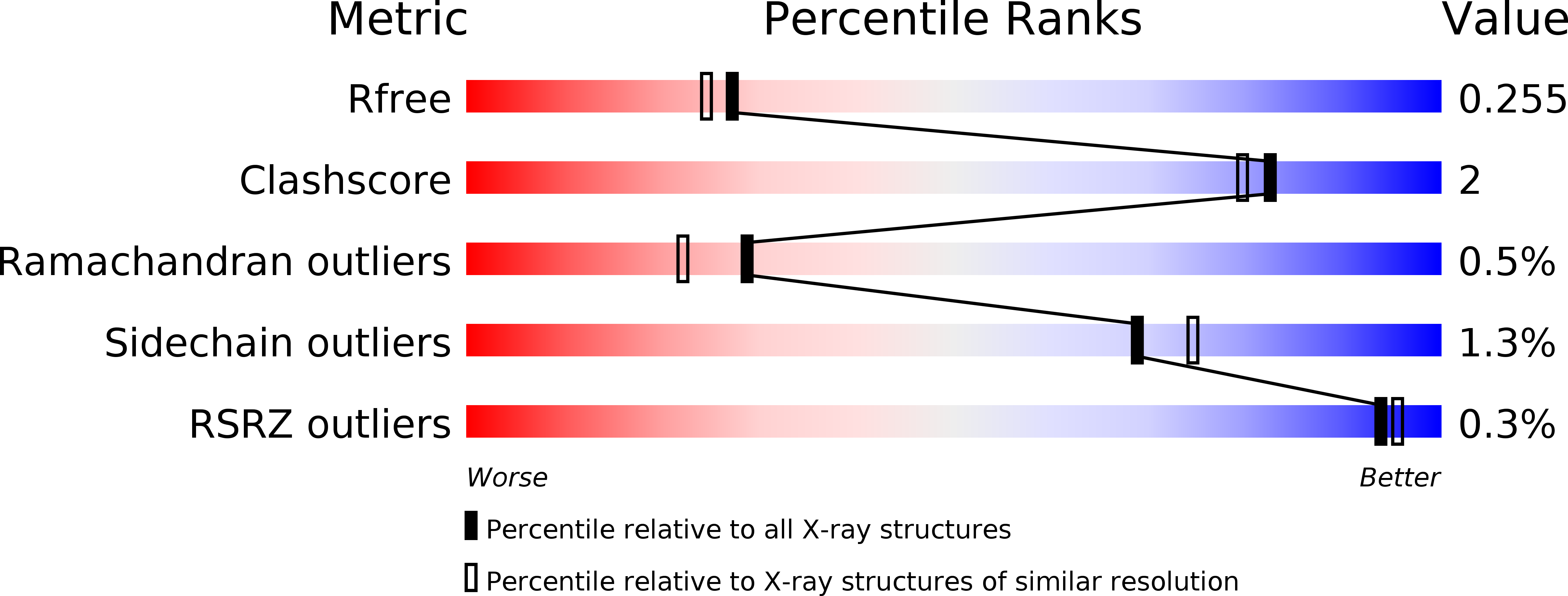
Resolution:
2.16 Å
R-Value Free:
0.25
R-Value Work:
0.19
R-Value Observed:
0.19
Space Group:
P 1
Macromolecular Entities
Polymer Type:polypeptide(L)
Description:Thymidylate kinase
Chain IDs:A (auth: B), B (auth: A)
Chain Length:195
Number of Molecules:2
Biological Source:Aquifex aeolicus (strain VF5)
Primary Citation
Structural studies of a hyperthermophilic thymidylate kinase enzyme reveal conformational substates along the reaction coordinate
FEBS J. 284 2527 2544 (2017)
PMID: 28627020 DOI: 10.1111/febs.14140

Abstact

UNLABELLED Thymidylate kinase (TMK) is a key enzyme which plays an important role in DNA synthesis. It belongs to the family of nucleoside monophosphate kinases, several of which undergo structure-encoded conformational changes to perform their function. However, the absence of three-dimensional structures for all the different reaction intermediates of a single TMK homolog hinders a clear understanding of its functional mechanism. We herein report the different conformational states along the reaction coordinate of a hyperthermophilic TMK from Aquifex aeolicus, determined via X-ray diffraction and further validated through normal-mode studies. The analyses implicate an arginine residue in the Lid region in catalysis, which was confirmed through site-directed mutagenesis and subsequent enzyme assays on the wild-type protein and mutants. Furthermore, the enzyme was found to exhibit broad specificity toward phosphate group acceptor nucleotides. Our comprehensive analyses of the conformational landscape of TMK, together with associated biochemical experiments, provide insights into the mechanistic details of TMK-driven catalysis, for example, the order of substrate binding and the reaction mechanism for phosphate transfer. Such a study has utility in the design of potent inhibitors for these enzymes. DATABASE Structural data are available in the PDB under the accession numbers 2PBR, 4S2E, 5H5B, 5XAI, 4S35, 5XB2, 5H56, 5XB3, 5H5K, 5XB5, and 5XBH.

Legend

Protein

Chemical

Disease

Primary Citation of related structures