Deposition Date
2017-03-15
Release Date
2017-08-02
Last Version Date
2024-03-27
Entry Detail
PDB ID:
5XAZ
Keywords:
Title:
Crystal structure of full length native tylp, a tetr regulator from streptomyces fradiae
Biological Source:
Source Organism(s):
Streptomyces fradiae (Taxon ID: 1906)
Expression System(s):
Method Details:
Experimental Method:
Resolution:
2.30 Å
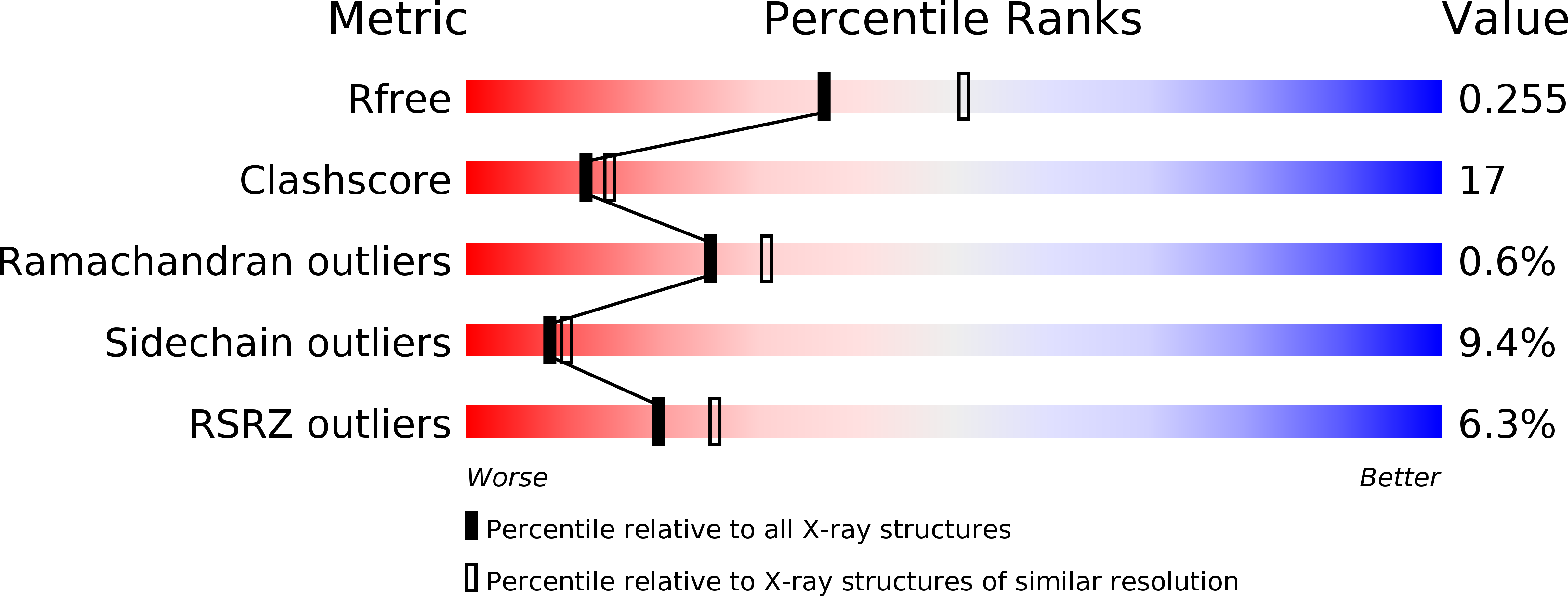
R-Value Free:
0.25
R-Value Work:
0.20
R-Value Observed:
0.20
Space Group:
P 1 21 1