
Deposition Date
2017-03-10
Release Date
2017-08-30
Last Version Date
2023-11-22
Entry Detail
PDB ID:
5X9Y
Keywords:
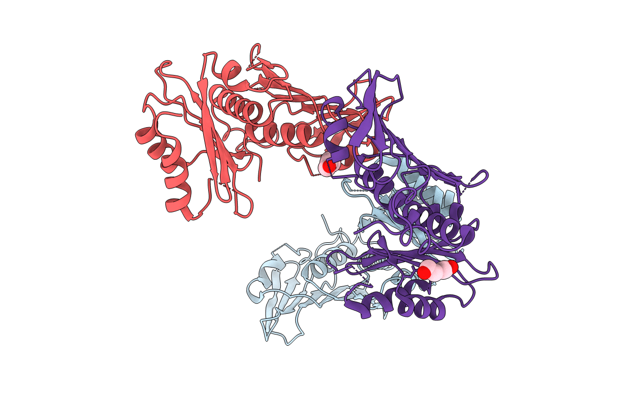
Title:
Crystal structure of the ATPase domain from bacterial mismatch repair endonuclease Aquifex aeolicus MutL.
Biological Source:
Source Organism:
Aquifex aeolicus (strain VF5) (Taxon ID: 224324)
Host Organism:
Method Details:
Experimental Method:
Resolution:
3.44 Å
R-Value Free:
0.29
R-Value Work:
0.25
R-Value Observed:
0.25
Space Group:
I 21 21 21
